Copyright
©2011 Baishideng Publishing Group Co.
World J Gastroenterol. Jun 21, 2011; 17(23): 2855-2859
Published online Jun 21, 2011. doi: 10.3748/wjg.v17.i23.2855
Published online Jun 21, 2011. doi: 10.3748/wjg.v17.i23.2855
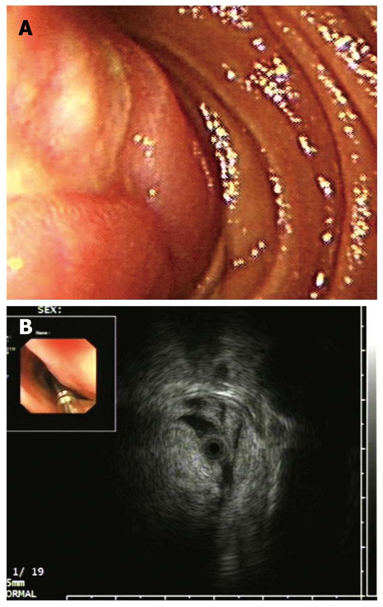
Figure 2 Endoscopic and endoscopic ultrasonography findings in Case 8.
A: A lobulated submucosal tumor in the descendant duodenum with an ulcer on its surface; B: Endoscopic ultrasonography showed an intensive hyperechoic lesion with a distinct anterior border originating from the submucosal layer without involvement of the overlying mucosal layer. The posterior border of the tumor was invisible because of the marked echo-attenuation.
- Citation: Chen HT, Xu GQ, Wang LJ, Chen YP, Li YM. Sonographic features of duodenal lipomas in eight clinicopathologically diagnosed patients. World J Gastroenterol 2011; 17(23): 2855-2859
- URL: https://www.wjgnet.com/1007-9327/full/v17/i23/2855.htm
- DOI: https://dx.doi.org/10.3748/wjg.v17.i23.2855


