Copyright
©2011 Baishideng Publishing Group Co.
World J Gastroenterol. Jun 7, 2011; 17(21): 2663-2666
Published online Jun 7, 2011. doi: 10.3748/wjg.v17.i21.2663
Published online Jun 7, 2011. doi: 10.3748/wjg.v17.i21.2663
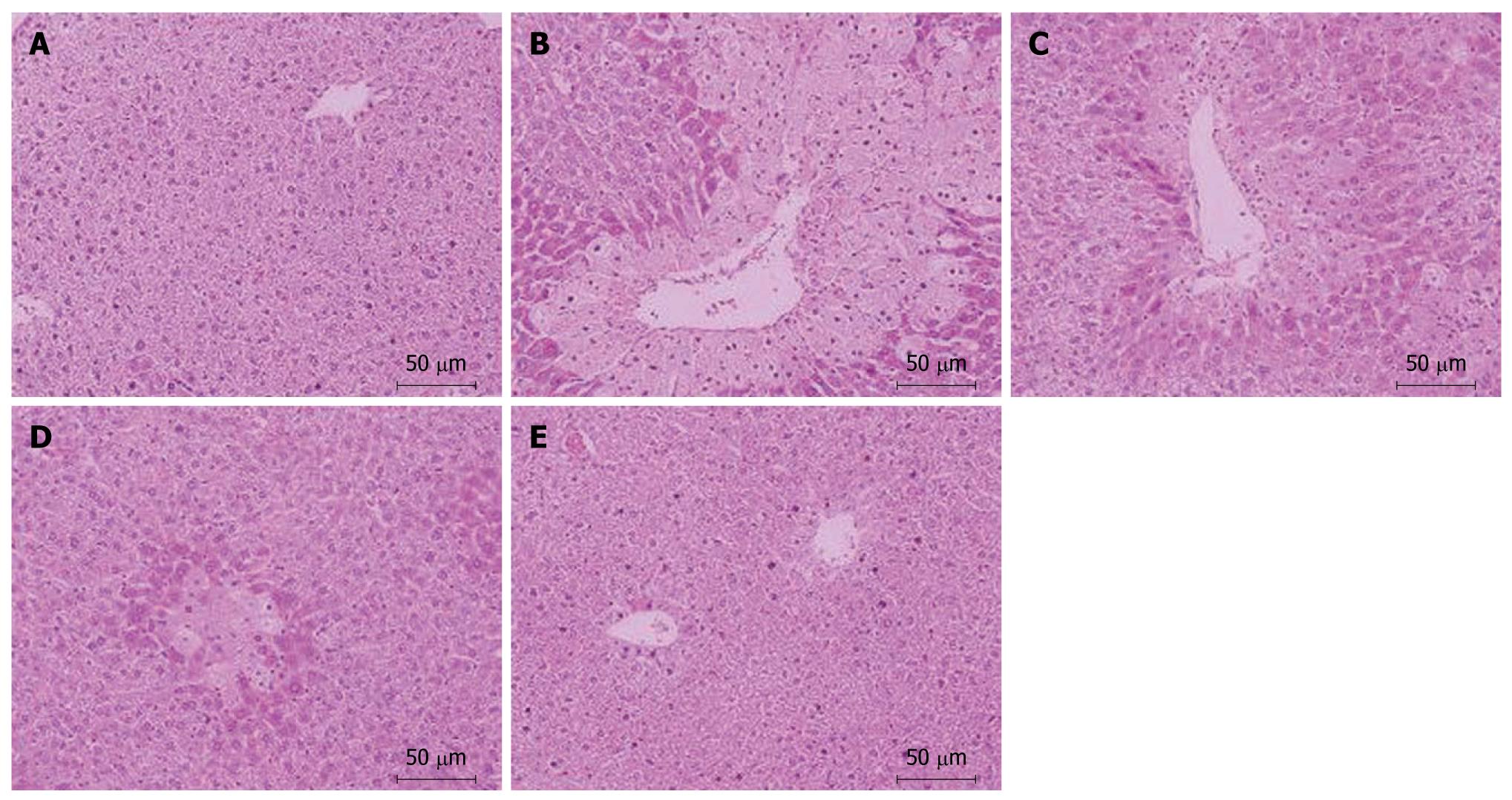
Figure 2 Liver histology of all groups 24 h after acetaminophen treatment (350 mg/kg).
Hematoxylin-eosin-stained liver sections displayed representative hepatocellular morphological changes. Original magnification × 200. A: Liver section in the vehicle control group showed a normal lobular structure; B: Liver section in acetaminophen alone group showed large areas of centrilobular necrosis, vacuolar degeneration and inflammatory cell infiltration; C: Liver section in the 200 mg/kg group showed necrosis with the same degree as in acetaminophen group; D: Liver section in the 400 mg/kg group showed a significant alleviation of liver injury; E: Liver section in the 800 mg/kg group showed absence of necrosis and almost normal lobular structure.
- Citation: He YY, Zhang BX, Jia FL. Protective effects of 2,4-dihydroxybenzophenone against acetaminophen-induced hepatotoxicity in mice. World J Gastroenterol 2011; 17(21): 2663-2666
- URL: https://www.wjgnet.com/1007-9327/full/v17/i21/2663.htm
- DOI: https://dx.doi.org/10.3748/wjg.v17.i21.2663


