Copyright
©2011 Baishideng Publishing Group Co.
World J Gastroenterol. Jun 7, 2011; 17(21): 2632-2640
Published online Jun 7, 2011. doi: 10.3748/wjg.v17.i21.2632
Published online Jun 7, 2011. doi: 10.3748/wjg.v17.i21.2632
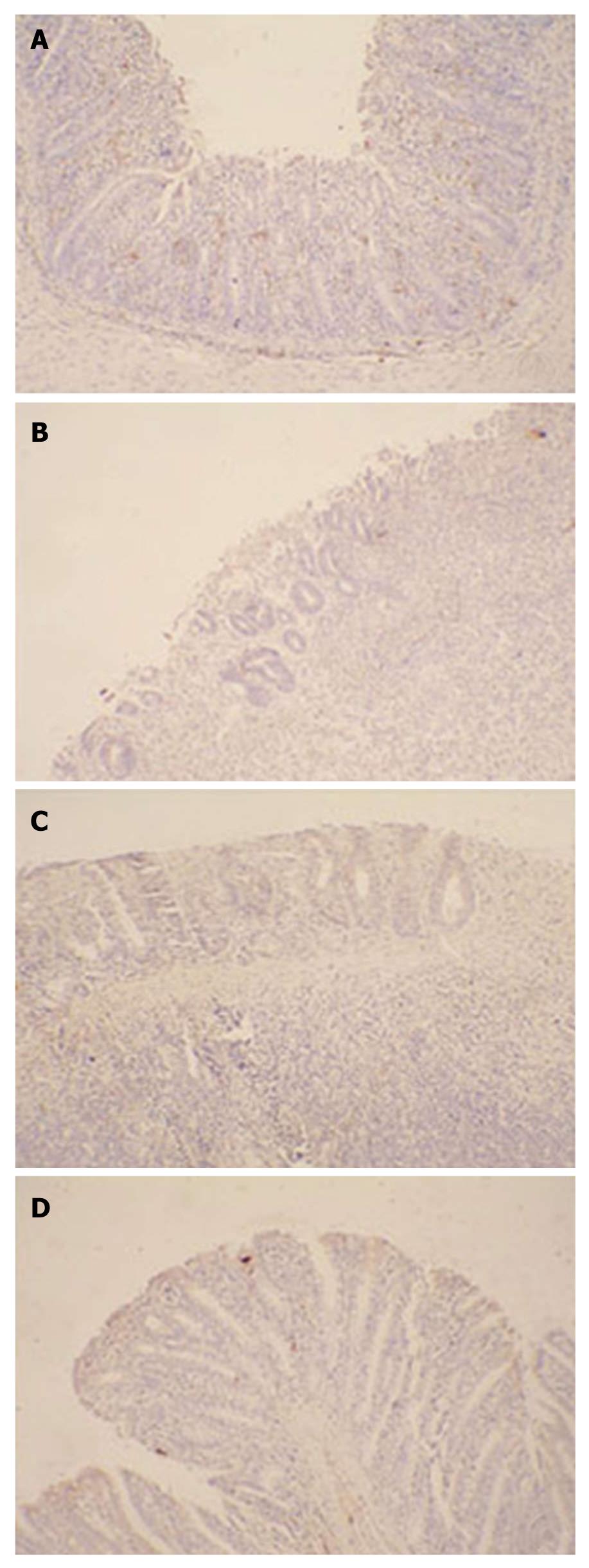
Figure 6 Immunohistochemistry of Ki67 in colon tissues.
The expression of the Ki67 protein was detected by immunohistochemistry with a rabbit anti-rat Ki67 antibody as the primary antibody and a horse radish peroxidase-conjugated goat anti-rabbit antibody as the secondary antibody, and the brown color was considered to be positive staining. The expression of Ki67 in damaged colonic tissues increased evidently in the group administered with attenuated Salmonella typhimurium Ty21a strain carrying human keratinocyte growth factor gene (SPK) strain, indicating the proliferation of colonic epithelial cells. A: Representative wound tissue from SPK group; B: Representative wound tissue from attenuated Salmonella typhimurium Ty21a strain group; C: Representative wound tissue from control group; D: Representative wound tissue from normal group (× 200).
- Citation: Liu CJ, Jin JD, Lv TD, Wu ZZ, Ha XQ. Keratinocyte growth factor gene therapy ameliorates ulcerative colitis in rats. World J Gastroenterol 2011; 17(21): 2632-2640
- URL: https://www.wjgnet.com/1007-9327/full/v17/i21/2632.htm
- DOI: https://dx.doi.org/10.3748/wjg.v17.i21.2632


