Copyright
©2011 Baishideng Publishing Group Co.
World J Gastroenterol. Jun 7, 2011; 17(21): 2632-2640
Published online Jun 7, 2011. doi: 10.3748/wjg.v17.i21.2632
Published online Jun 7, 2011. doi: 10.3748/wjg.v17.i21.2632
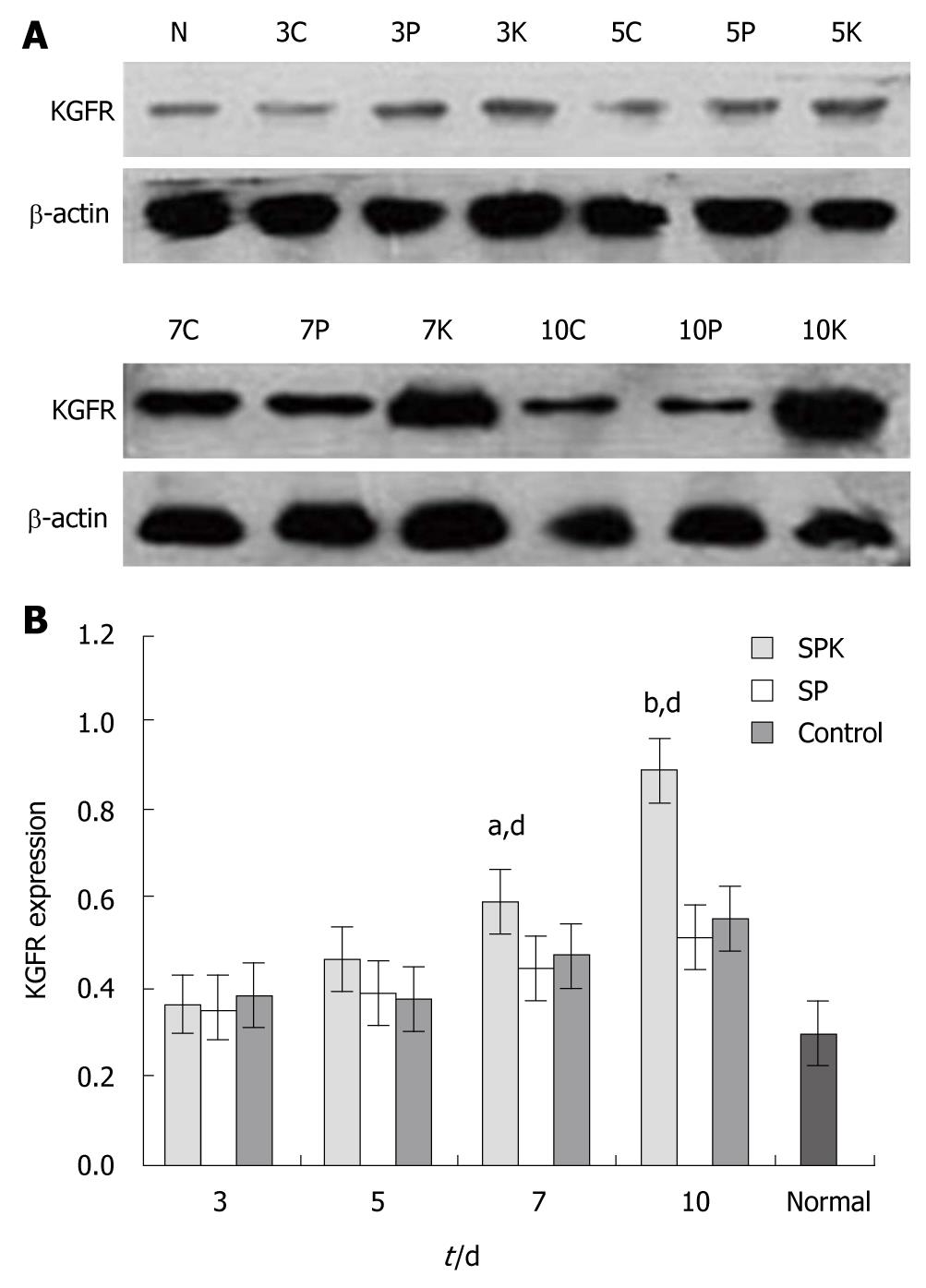
Figure 4 Western blotting of keratinocyte growth factor receptor in colon tissues.
The expression of keratinocyte growth factor receptor (KGFR) in the colon tissues was measured by Western blotting after drug administration. The expression of KGFR on days 7 and 10 increased obviously in attenuated Salmonella typhimurium Ty21a strain carrying human keratinocyte growth factor gene group (SPK) compared with attenuated Salmonella typhimurium Ty21a strain (SP) and control groups. A: Western blotting analysis of KGFR. N: normal group; 3C, 3P and 3K represents control, SP and SPK groups, respectively, on day 3 of drug administration; 5C, 5P and 5K, on day 5; 7C, 7P and 7K, on day 7; 10 C, 10P and 10K, on day 10; B: Analysis of Western blotting of KGFR. The density of the bands was quantified using image-pro plus 6.0 software, and the data are presented as mean ± SD, n = 6. aP < 0.05, bP < 0.01 vs control group; dP < 0.01, vs SP group at the same time point.
- Citation: Liu CJ, Jin JD, Lv TD, Wu ZZ, Ha XQ. Keratinocyte growth factor gene therapy ameliorates ulcerative colitis in rats. World J Gastroenterol 2011; 17(21): 2632-2640
- URL: https://www.wjgnet.com/1007-9327/full/v17/i21/2632.htm
- DOI: https://dx.doi.org/10.3748/wjg.v17.i21.2632


