Copyright
©2011 Baishideng Publishing Group Co.
World J Gastroenterol. Jun 7, 2011; 17(21): 2602-2610
Published online Jun 7, 2011. doi: 10.3748/wjg.v17.i21.2602
Published online Jun 7, 2011. doi: 10.3748/wjg.v17.i21.2602
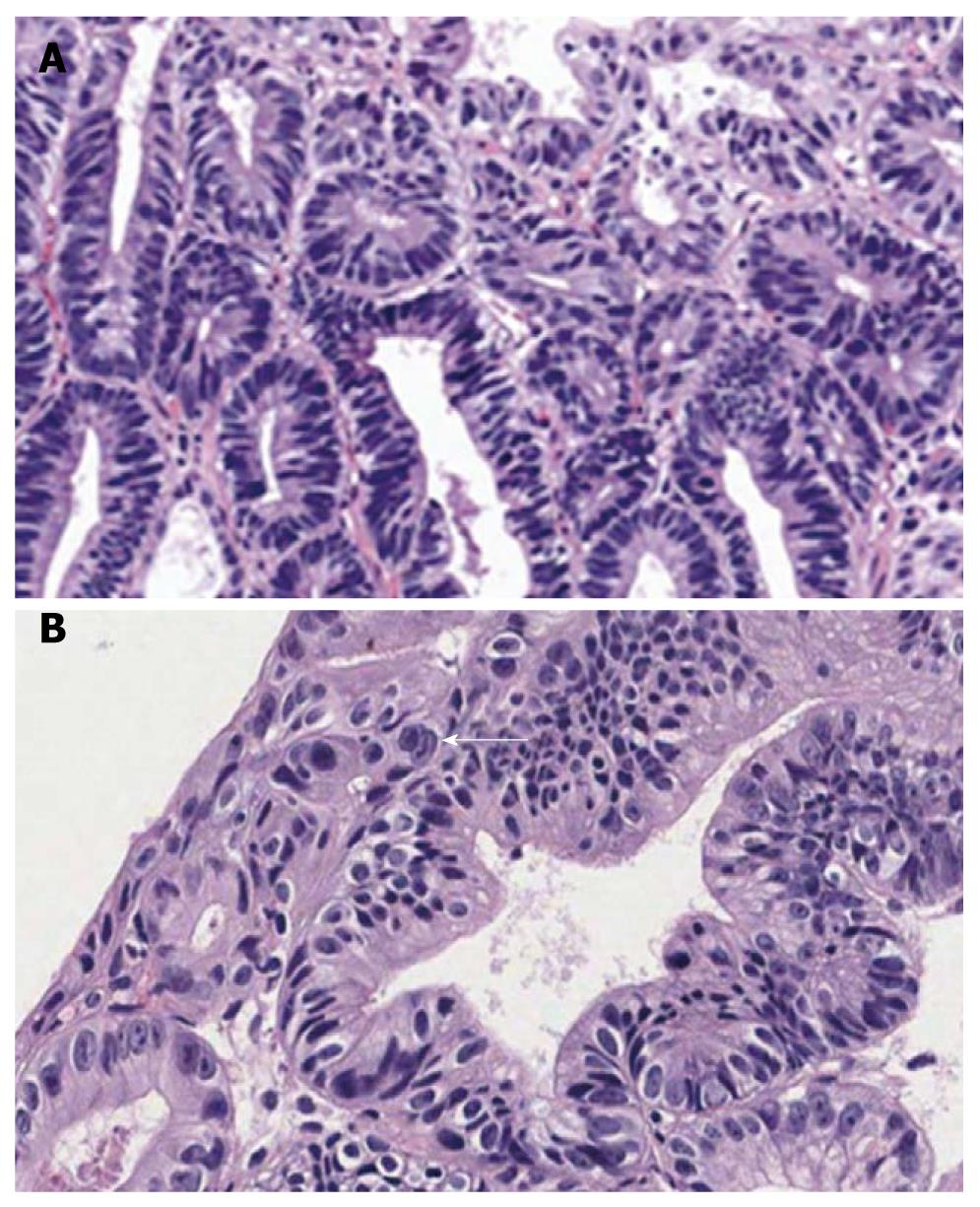
Figure 7 Major diagnosis after the consensus conference of adenocarcinoma.
A: Compact small glandular proliferation with budding and branching. Regular glandular size and distribution (HE, × 100); B: Severe nuclear stratification approaching the top of the cytoplasm in more than three contiguous glands. Marked hyperchromasia and mitoses with invasion into the lamina propria (arrow) (HE, × 400).
- Citation: Kim JM, Cho MY, Sohn JH, Kang DY, Park CK, Kim WH, Jin SY, Kim KM, Chang HK, Yu E, Jung ES, Chang MS, Joo JE, Joo M, Kim YW, Park DY, Kang YK, Park SH, Han HS, Kim YB, Park HS, Chae YS, Kwon KW, Chang HJ, Pathologists TGPSGOKSO. Diagnosis of gastric epithelial neoplasia: Dilemma for Korean pathologists. World J Gastroenterol 2011; 17(21): 2602-2610
- URL: https://www.wjgnet.com/1007-9327/full/v17/i21/2602.htm
- DOI: https://dx.doi.org/10.3748/wjg.v17.i21.2602


