Copyright
©2011 Baishideng Publishing Group Co.
World J Gastroenterol. May 21, 2011; 17(19): 2397-2406
Published online May 21, 2011. doi: 10.3748/wjg.v17.i19.2397
Published online May 21, 2011. doi: 10.3748/wjg.v17.i19.2397
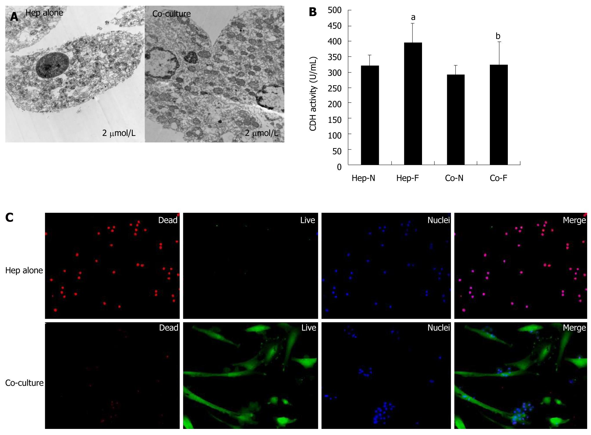
Figure 4 Cytotoxic effects of liver failure serum on hepatocytes cultured alone vs co-cultured hepatocytes.
A: Transmission electron microscopy (TEM) analysis of the homo-cultured hepatocytes (left panel) and hepatocytes co-cultured with mesenchymal stem cells (MSCs) (righ panel) in the presence of 60% of liver failure serum; B: Lactate dehydrogenase (LDH) release assay of hepatocytes was indicated in each group; C: Live/dead assay of homo-cultured hepatocytes (upper panel) and hepatocytes co-cultured with MSCs (lower panel) in the presence of 60% of liver failure serum. aIndicates significant difference vs Hep-N group; bIndicates significant difference vs homo-hepatocytes cultured by various concentrations of liver failure serum (Hep-F) group. Hep-N: Hepatocytes homo-cultured with normal serum; Co-N:Hepatocytes co-cultured with normal serum; Co-F: Liver failure serum.
-
Citation: Shi XL, Gu JY, Zhang Y, Han B, Xiao JQ, Yuan XW, Zhang N, Ding YT. Protective effects of ACLF sera on metabolic functions and proliferation of hepatocytes co-cultured with bone marrow MSCs
in vitro . World J Gastroenterol 2011; 17(19): 2397-2406 - URL: https://www.wjgnet.com/1007-9327/full/v17/i19/2397.htm
- DOI: https://dx.doi.org/10.3748/wjg.v17.i19.2397


