Copyright
©2011 Baishideng Publishing Group Co.
World J Gastroenterol. May 21, 2011; 17(19): 2379-2388
Published online May 21, 2011. doi: 10.3748/wjg.v17.i19.2379
Published online May 21, 2011. doi: 10.3748/wjg.v17.i19.2379
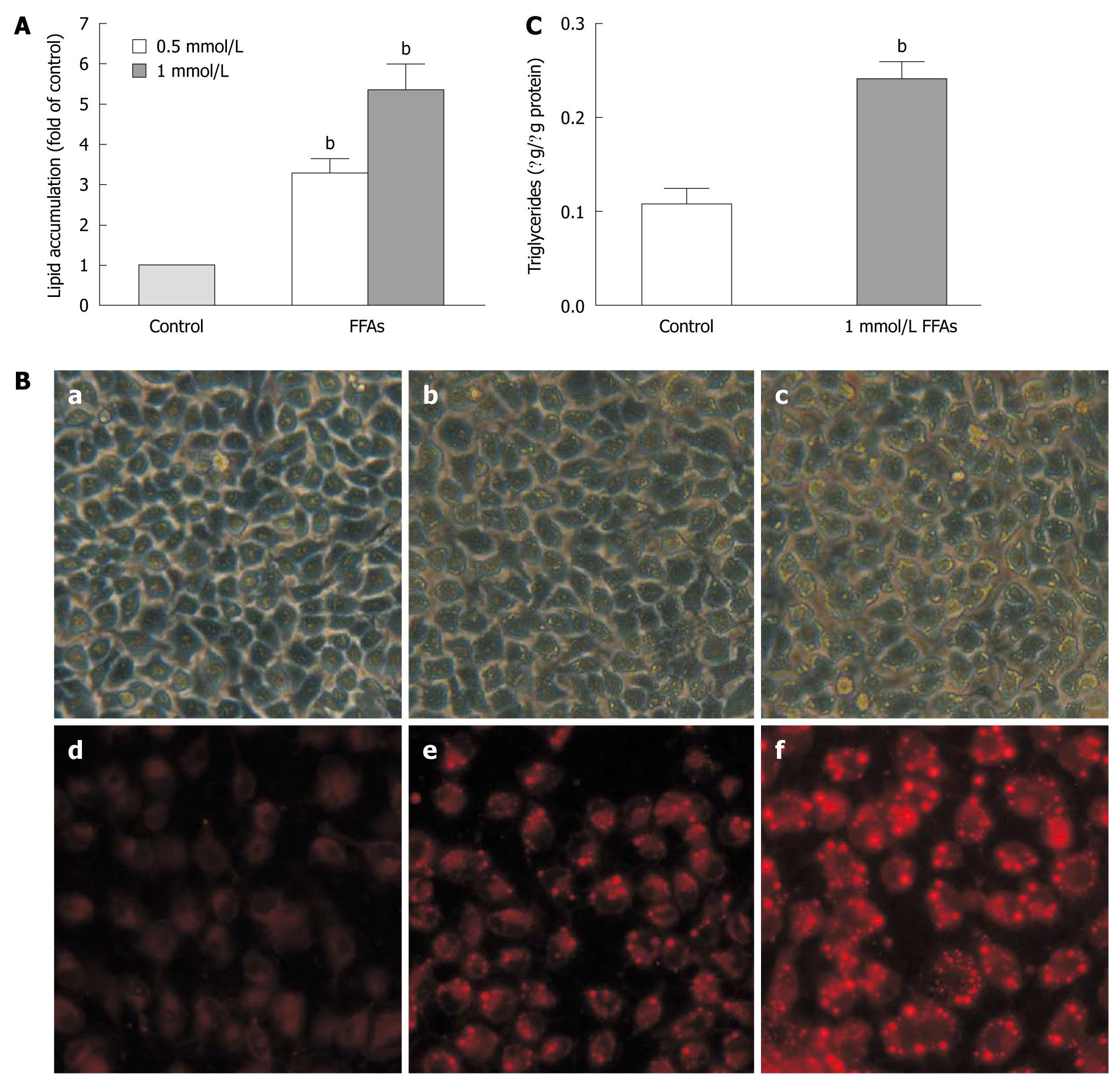
Figure 3 Free fatty acid induced lipid accumulation in L-02 cells.
A: L-02 cells were incubated with a free fatty acid (FFA) mixture (oleate and palmitate at the ratio of 2:1) for 24 h. Intracellular lipid accumulation was evaluated after Nile red staining. Results were expressed as mean ± SE of three independent experiments. bP < 0.01 vs control group; B: Representative micrographs showing intracellular lipid accumulation in L-02 cells as observed by phase-contrast microscopy (panels a-c) and fluorescence microscopy (panels d-f). Panels a/d, b/e and c/f are control cells, cells treated with 0.5 and 1 mmol/L FFA, respectively; C: Triglyceride levels in L-02 cells treated with 1 mmol/L FFA. Results were expressed as mean ± SE of three independent experiments. bP < 0.01 vs control group.
- Citation: Chu JH, Wang H, Ye Y, Chan PK, Pan SY, Fong WF, Yu ZL. Inhibitory effect of schisandrin B on free fatty acid-induced steatosis in L-02 cells. World J Gastroenterol 2011; 17(19): 2379-2388
- URL: https://www.wjgnet.com/1007-9327/full/v17/i19/2379.htm
- DOI: https://dx.doi.org/10.3748/wjg.v17.i19.2379


