Copyright
©2011 Baishideng Publishing Group Co.
World J Gastroenterol. May 21, 2011; 17(19): 2379-2388
Published online May 21, 2011. doi: 10.3748/wjg.v17.i19.2379
Published online May 21, 2011. doi: 10.3748/wjg.v17.i19.2379
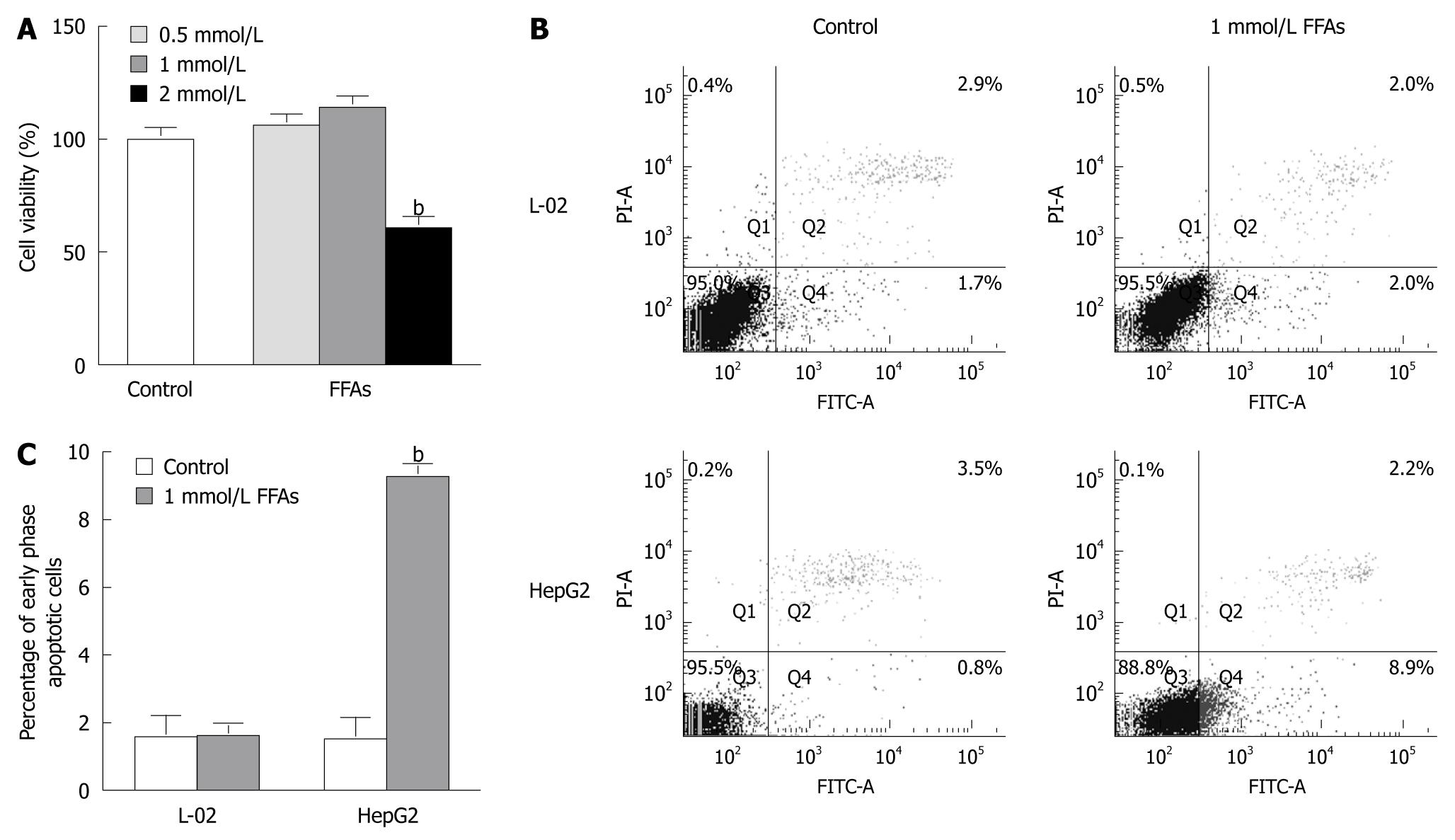
Figure 2 Cytotoxic and apoptotic effects of free fatty acid treatment on cultured cells.
A: L-02 cells were treated with a free fatty acid (FFA) mixture (oleate and palmitate at the ratio of 2:1) at various concentrations for 24 h. Cell viability was determined by the 3-(4,5)-dimethylthiahiazo (-z-y1)-3,5-di- phenytetrazoliumromide (MTT) assay. bP < 0.01 vs control group; B: L-02 and HepG2 cells were treated with 1 mmol/L FFA mixture (oleate and palmitate at the ratio of 2:1) for 24 h and stained with Annexin V-fluorescein isothiocyanate (FITC) and propidium iodide. Apoptotic and necrotic cells were monitored by flow cytometry. Normal, early and late apoptotic cells as well as necrotic cells were shown in Q3, Q4, Q2 and Q1 quadrants, respectively. The percentage of cells in each quadrant was displayed. Results were the representative of three independent experiments; C: Quantification of early phase apoptotic cells in response to FFA treatment. bP < 0.01 vs HepG2 control group.
- Citation: Chu JH, Wang H, Ye Y, Chan PK, Pan SY, Fong WF, Yu ZL. Inhibitory effect of schisandrin B on free fatty acid-induced steatosis in L-02 cells. World J Gastroenterol 2011; 17(19): 2379-2388
- URL: https://www.wjgnet.com/1007-9327/full/v17/i19/2379.htm
- DOI: https://dx.doi.org/10.3748/wjg.v17.i19.2379


