Copyright
©2011 Baishideng Publishing Group Co.
World J Gastroenterol. May 14, 2011; 17(18): 2332-2337
Published online May 14, 2011. doi: 10.3748/wjg.v17.i18.2332
Published online May 14, 2011. doi: 10.3748/wjg.v17.i18.2332
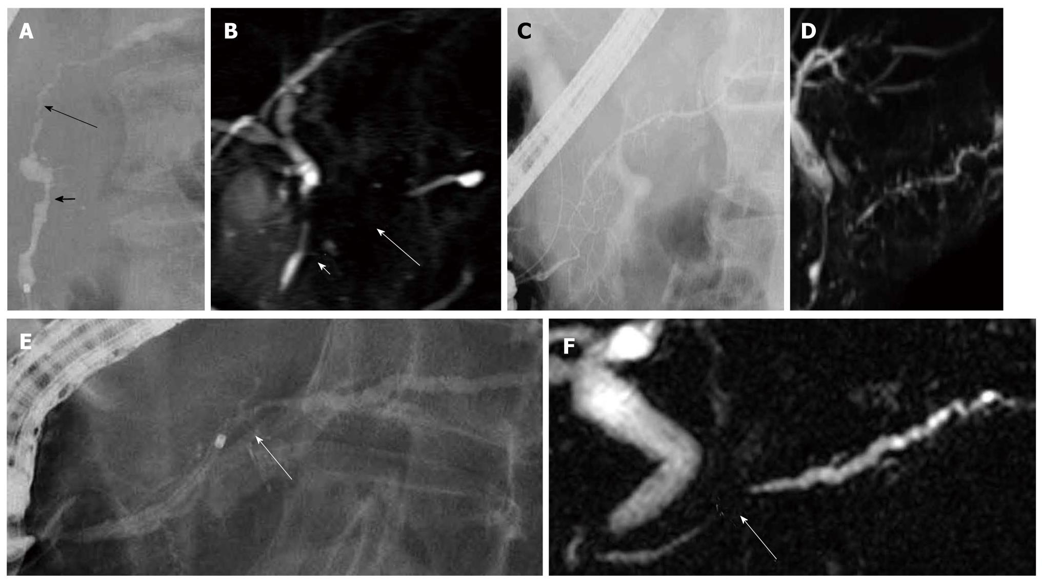
Figure 1 Pancreatography of autoimmune pancreatitis.
A: Endoscopic retrograde cholangiopancreatography finding of autoimmune pancreatitis showing skipped lesions of the main pancreatic duct (short and long arrows); B: On magnetic resonance cholangiopancreatography, skipped lesions (short and long arrows) on endoscopic retrograde cholangiopancreatography were not visualized; C: Endoscopic retrograde cholangiopancreatography finding of autoimmune pancreatitis showing side branch derivation from the narrowed portion of the main pancreatic duct; D: Recent magnetic resonance cholangiopancreatography could show diffuse narrowing of the main pancreatic duct fairly well; E: Endoscopic retrograde cholangiopancreatography finding of autoimmune pancreatitis showing a short narrowed main pancreatic duct (arrow) with upstream dilatation less than 5 mm; F: On magnetic resonance cholangiopancreatography, the narrowed portion (arrow) was not visualized.
- Citation: Takuma K, Kamisawa T, Tabata T, Inaba Y, Egawa N, Igarashi Y. Utility of pancreatography for diagnosing autoimmune pancreatitis. World J Gastroenterol 2011; 17(18): 2332-2337
- URL: https://www.wjgnet.com/1007-9327/full/v17/i18/2332.htm
- DOI: https://dx.doi.org/10.3748/wjg.v17.i18.2332


