Copyright
©2011 Baishideng Publishing Group Co.
World J Gastroenterol. May 7, 2011; 17(17): 2255-2258
Published online May 7, 2011. doi: 10.3748/wjg.v17.i17.2255
Published online May 7, 2011. doi: 10.3748/wjg.v17.i17.2255
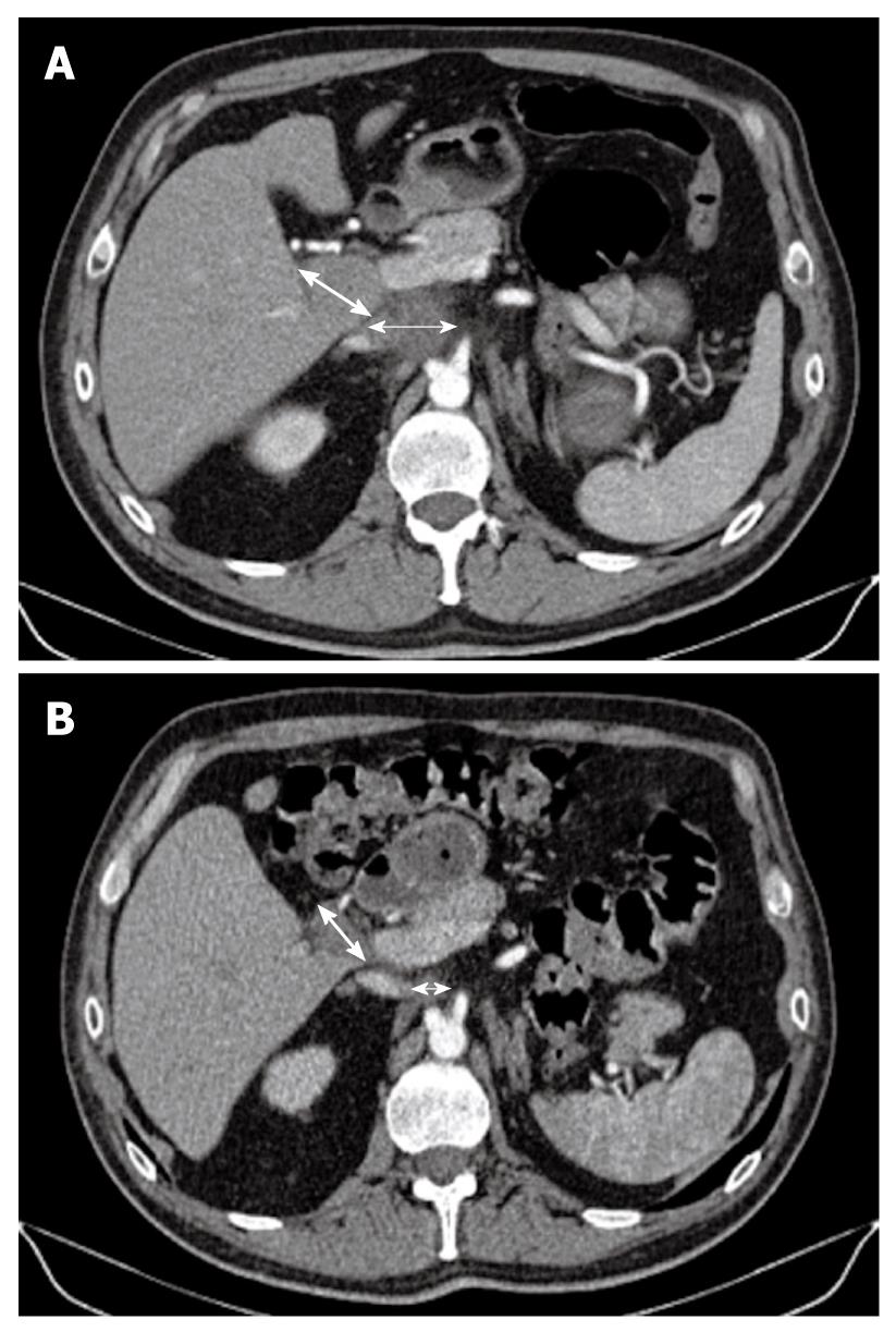
Figure 2 Computed tomography scan.
A: Initially, there were two metastatic lymph nodes (MLNs): one celiac (thin arrow) measuring 4 cm and one measuring 3 cm located in the liver hilum (thick arrow); B: Computed tomography scan taken after the end of the eight cycles of gemcitabine plus oxaliplatin combined with sorafenib, showed that the MLNs (thin arrow for celiac node and thick arrow for hilar node) decreased in size dramatically with a more necrotic aspect, which allowed secondary surgery followed by radiotherapy.
- Citation: Williet N, Dubreuil O, Boussaha T, Trouilloud I, Landi B, Housset M, Botti M, Rougier P, Belghiti J, Taieb J. Neoadjuvant sorafenib combined with gemcitabine plus oxaliplatin in advanced hepatocellular carcinoma. World J Gastroenterol 2011; 17(17): 2255-2258
- URL: https://www.wjgnet.com/1007-9327/full/v17/i17/2255.htm
- DOI: https://dx.doi.org/10.3748/wjg.v17.i17.2255


