Copyright
©2011 Baishideng Publishing Group Co.
World J Gastroenterol. Apr 21, 2011; 17(15): 1947-1960
Published online Apr 21, 2011. doi: 10.3748/wjg.v17.i15.1947
Published online Apr 21, 2011. doi: 10.3748/wjg.v17.i15.1947
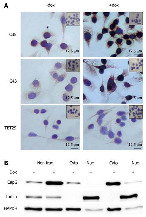
Figure 7 Subcellular localization of CapG in stable CapG inducible clones by immunohistochemistry (A) and subcellular fractionation (B).
A: CapG protein expression and localization in Sβtet29Cap35 (C35), Sβtet29Cap43 (C43) and the control Sβtet29 (TET29) cells was analyzed 24 h after treatment with 500 ng/ml doxycycline (+ dox) or PBS as control (- dox). Specific antibody reaction (anti-CapG) was visualized by a peroxidase labelled secondary antibody (DAB detection, brown colour). Nuclei were counterstained with hematoxylin (blue colour). Bars = 12.5 μm. B. Whole protein lysate (non frac.) or enriched nuclear (Nuc) and cytoplasmic (Cyto) fractions of Sβtet29Cap35, Sβtet29Cap43 and the control Sβtet29 clones 24 h after treatment with 500 ng/mL doxycycline (+) or PBS as control (-) were analysed using Western blotting for the detection of CapG, Lamin (marker for nuclear fraction) and GAPDH (marker for cytoplasmic fraction). The experiment was performed three times. A representative Western blot of Sβtet29Cap35 is shown.
- Citation: Tonack S, Patel S, Jalali M, Nedjadi T, Jenkins RE, Goldring C, Neoptolemos J, Costello E. Tetracycline-inducible protein expression in pancreatic cancer cells: Effects of CapG overexpression. World J Gastroenterol 2011; 17(15): 1947-1960
- URL: https://www.wjgnet.com/1007-9327/full/v17/i15/1947.htm
- DOI: https://dx.doi.org/10.3748/wjg.v17.i15.1947


