Copyright
©2011 Baishideng Publishing Group Co.
World J Gastroenterol. Apr 14, 2011; 17(14): 1923-1926
Published online Apr 14, 2011. doi: 10.3748/wjg.v17.i14.1923
Published online Apr 14, 2011. doi: 10.3748/wjg.v17.i14.1923
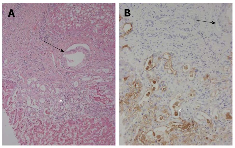
Figure 4 Bileductular cell proliferation.
A: Clusters of atypical bile ductular cells (*) are seen, and are regarded as malignant cells. An arrow shows the involvement of carcinoma cells of interlobular bile ducts. Hematoxylin and eosin staining (H and E); B: Atypical bile ductular cells (*) are positive for mucin (MUC)1. Neoplastic epithelial cells replacing interlobular bile ducts (arrow) are also focally positive. Immunostaining for MUC1 and hematoxylin.
- Citation: Xu J, Sato Y, Harada K, NorihideYoneda, Ueda T, Kawashima A, AkishiOoi, YasuniNakanuma. Intraductal papillary neoplasm of the bile duct in liver cirrhosis with hepatocellular carcinoma. World J Gastroenterol 2011; 17(14): 1923-1926
- URL: https://www.wjgnet.com/1007-9327/full/v17/i14/1923.htm
- DOI: https://dx.doi.org/10.3748/wjg.v17.i14.1923


