Copyright
©2011 Baishideng Publishing Group Co.
World J Gastroenterol. Apr 14, 2011; 17(14): 1923-1926
Published online Apr 14, 2011. doi: 10.3748/wjg.v17.i14.1923
Published online Apr 14, 2011. doi: 10.3748/wjg.v17.i14.1923
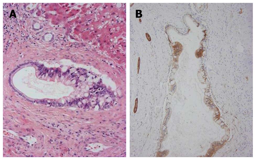
Figure 3 Carcinomatouscholangiocytes.
A: Atypical biliary epithelial cells (cholangiocarcinoma) partially replace the epithelia of septal bile ducts. Hematoxylin and eosin staining (H and E); B: Neoplastic biliary epithelial cells spreading on the luminal surface of the septal bile ducts are positive for neural cell adhesion molecule (NCAM). Nerve fibers around the bile ducts are also positive. Immunostaining for NCAM and hematoxylin.
- Citation: Xu J, Sato Y, Harada K, NorihideYoneda, Ueda T, Kawashima A, AkishiOoi, YasuniNakanuma. Intraductal papillary neoplasm of the bile duct in liver cirrhosis with hepatocellular carcinoma. World J Gastroenterol 2011; 17(14): 1923-1926
- URL: https://www.wjgnet.com/1007-9327/full/v17/i14/1923.htm
- DOI: https://dx.doi.org/10.3748/wjg.v17.i14.1923


