Copyright
©2011 Baishideng Publishing Group Co.
World J Gastroenterol. Apr 14, 2011; 17(14): 1923-1926
Published online Apr 14, 2011. doi: 10.3748/wjg.v17.i14.1923
Published online Apr 14, 2011. doi: 10.3748/wjg.v17.i14.1923
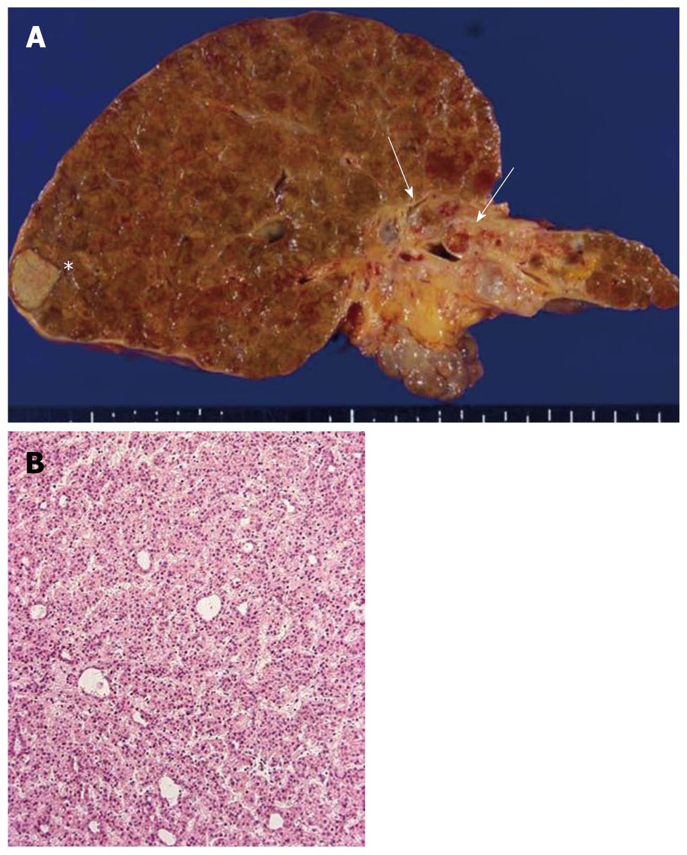
Figure 1 Liver pathology.
A: The autopsied liver tissue shows cirrhosis with a solid whitish lesion (*) in segment 6 of the right lobe (hepatocellular carcinoma after transcatheter arterial chemoembolization) and cystic dilatation in the left intrahepatic bile ducts filling with yellowish-tan papillary masses and mucin production (arrows). The left liver appears atrophied; B: Well-differentiated hepatocellular carcinoma. Hematoxylin and eosin staining (H and E).
- Citation: Xu J, Sato Y, Harada K, NorihideYoneda, Ueda T, Kawashima A, AkishiOoi, YasuniNakanuma. Intraductal papillary neoplasm of the bile duct in liver cirrhosis with hepatocellular carcinoma. World J Gastroenterol 2011; 17(14): 1923-1926
- URL: https://www.wjgnet.com/1007-9327/full/v17/i14/1923.htm
- DOI: https://dx.doi.org/10.3748/wjg.v17.i14.1923


