Copyright
©2011 Baishideng Publishing Group Co.
World J Gastroenterol. Apr 7, 2011; 17(13): 1701-1709
Published online Apr 7, 2011. doi: 10.3748/wjg.v17.i13.1701
Published online Apr 7, 2011. doi: 10.3748/wjg.v17.i13.1701
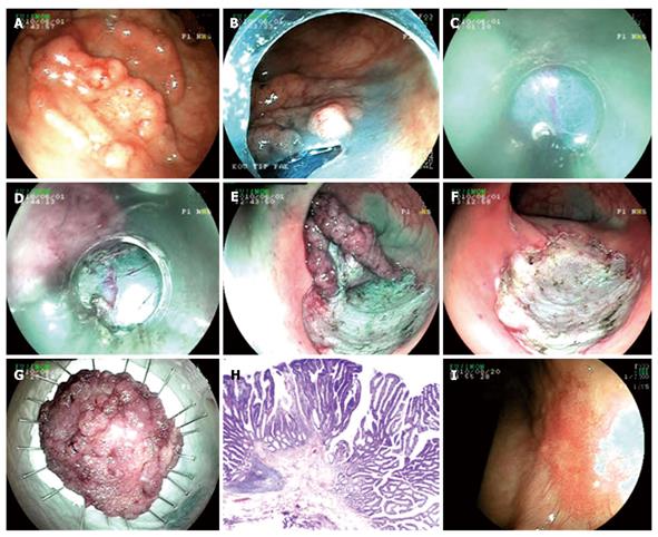
Figure 4 Endoscopic submucosal dissection procedure for pseudo-depressed type lateral spreading tumor with high grade dysplasia at rectum.
A: Pseudo-depressed type lateral spreading tumor at rectum; B: Cutting the lesion circumferentially with endo-cut; C, D: Submucosal dissection with semipermeable cap; E: Endoscopic view just before completing submucosal dissection; F: Appearance of the mucosa after the lesion being extracted; G: Microscopic view of the lesion; H: Histology; tubulovillous adenoma including fields of focal pattern loss and dysplasia (HE × 20); I: Endoscopic view ten weeks after the procedure.
- Citation: Hulagu S, Senturk O, Aygun C, Kocaman O, Celebi A, Konduk T, Koc D, Sirin G, Korkmaz U, Duman AE, Bozkurt N, Dindar G, Attila T, Gurbuz Y, Tarcin O, Kalayci C. Endoscopic submucosal dissection for premalignant lesions and noninvasive early gastrointestinal cancers. World J Gastroenterol 2011; 17(13): 1701-1709
- URL: https://www.wjgnet.com/1007-9327/full/v17/i13/1701.htm
- DOI: https://dx.doi.org/10.3748/wjg.v17.i13.1701


