Copyright
©2011 Baishideng Publishing Group Co.
World J Gastroenterol. Apr 7, 2011; 17(13): 1701-1709
Published online Apr 7, 2011. doi: 10.3748/wjg.v17.i13.1701
Published online Apr 7, 2011. doi: 10.3748/wjg.v17.i13.1701
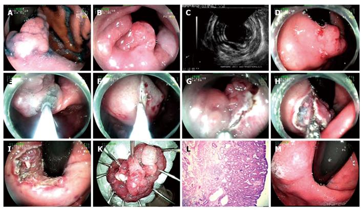
Figure 2 Endoscopic submucosal dissection procedure for adenoma with high grade dysplasia at cardia.
A: Adenoma at cardia; B: View of the lesion from esophageal aspect; C: Endosonographic image of the lesion; D, E: Marking the borders of the lesion with needle knife and lifting it; F, G: Cutting the lesion circumferentially with endo-cut above Z line, in retroflexion; H: Dissection of the submucosal area; I: Appearance of the mucosa after the lesion being extracted; K: Microscopic view of the lesion; L: Histology: mucosa, muscularis mucosa and superficial submucosa of stomach (HE × 20). Adenoma structure including adenomatous epithelium formed by irregular glands at mucosa; M: Endoscopic view six months after the procedure.
- Citation: Hulagu S, Senturk O, Aygun C, Kocaman O, Celebi A, Konduk T, Koc D, Sirin G, Korkmaz U, Duman AE, Bozkurt N, Dindar G, Attila T, Gurbuz Y, Tarcin O, Kalayci C. Endoscopic submucosal dissection for premalignant lesions and noninvasive early gastrointestinal cancers. World J Gastroenterol 2011; 17(13): 1701-1709
- URL: https://www.wjgnet.com/1007-9327/full/v17/i13/1701.htm
- DOI: https://dx.doi.org/10.3748/wjg.v17.i13.1701


