Copyright
©2011 Baishideng Publishing Group Co.
World J Gastroenterol. Apr 7, 2011; 17(13): 1701-1709
Published online Apr 7, 2011. doi: 10.3748/wjg.v17.i13.1701
Published online Apr 7, 2011. doi: 10.3748/wjg.v17.i13.1701
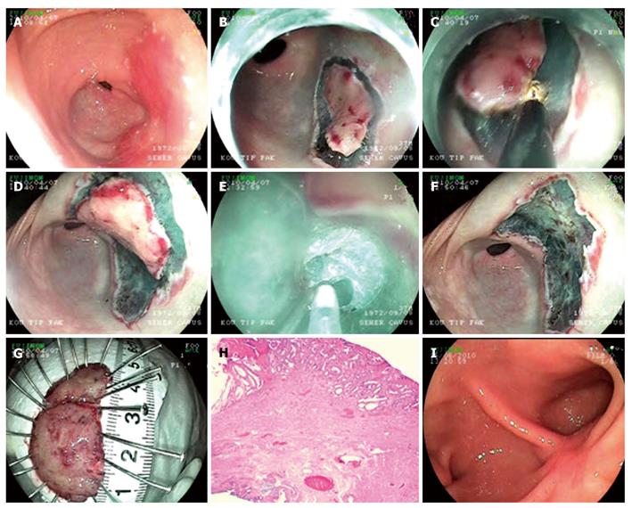
Figure 1 Endoscopic submucosal dissection procedure for adenoma with high grade dysplasia at antrum.
A: Endoscopic view flat adenoma at antrum; B: Cutting of the first piece of the lesion which was decided to be extracted in two pieces; C: Submucosal dissection of the first piece; D: Cutting of the second piece of the lesion; E: Submucosal dissection of the second piece; F: Endoscopic view after the lesion is being extracted; G: Microscopic view of the lesion; H: Histology; Adenoma including fields of marked glandular atypia and distortion (HE × 20); I: Endoscopic view ten weeks after the procedure.
- Citation: Hulagu S, Senturk O, Aygun C, Kocaman O, Celebi A, Konduk T, Koc D, Sirin G, Korkmaz U, Duman AE, Bozkurt N, Dindar G, Attila T, Gurbuz Y, Tarcin O, Kalayci C. Endoscopic submucosal dissection for premalignant lesions and noninvasive early gastrointestinal cancers. World J Gastroenterol 2011; 17(13): 1701-1709
- URL: https://www.wjgnet.com/1007-9327/full/v17/i13/1701.htm
- DOI: https://dx.doi.org/10.3748/wjg.v17.i13.1701


