Copyright
©2011 Baishideng Publishing Group Co.
World J Gastroenterol. Mar 28, 2011; 17(12): 1649-1654
Published online Mar 28, 2011. doi: 10.3748/wjg.v17.i12.1649
Published online Mar 28, 2011. doi: 10.3748/wjg.v17.i12.1649
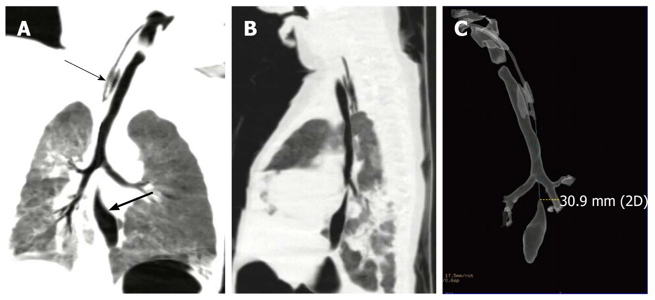
Figure 1 The reconstruction techniques of multidetector-row computed tomography show a case of esophageal atresia and tracheoesophageal fistula with a long inter-pouch gap.
A: Coronal view of multiple planar volume reconstruction showing a long gap between the proximal pouch (thin arrow) and distal pouch (thick arrow) as well as the tracheobronchial tree with an aspirating catheter in the proximal esophageal pouch; B: Oblique sagittal view of multiple planar volume reconstruction showing a long inter-pouch distance with a tenuous fistula; C: Posteroanterior projection of transparency lung volume rendering demonstrating the distance between esophageal segments and three-dimensional anatomy of esophageal pouches and tracheobronchial tree after removal of lungs in case 3 (a 3-d old male neonate).
- Citation: Wen Y, Peng Y, Zhai RY, Li YZ. Application of MPVR and TL-VR with 64-row MDCT in neonates with congenital EA and distal TEF. World J Gastroenterol 2011; 17(12): 1649-1654
- URL: https://www.wjgnet.com/1007-9327/full/v17/i12/1649.htm
- DOI: https://dx.doi.org/10.3748/wjg.v17.i12.1649


