Copyright
©2011 Baishideng Publishing Group Co.
World J Gastroenterol. Mar 28, 2011; 17(12): 1584-1593
Published online Mar 28, 2011. doi: 10.3748/wjg.v17.i12.1584
Published online Mar 28, 2011. doi: 10.3748/wjg.v17.i12.1584
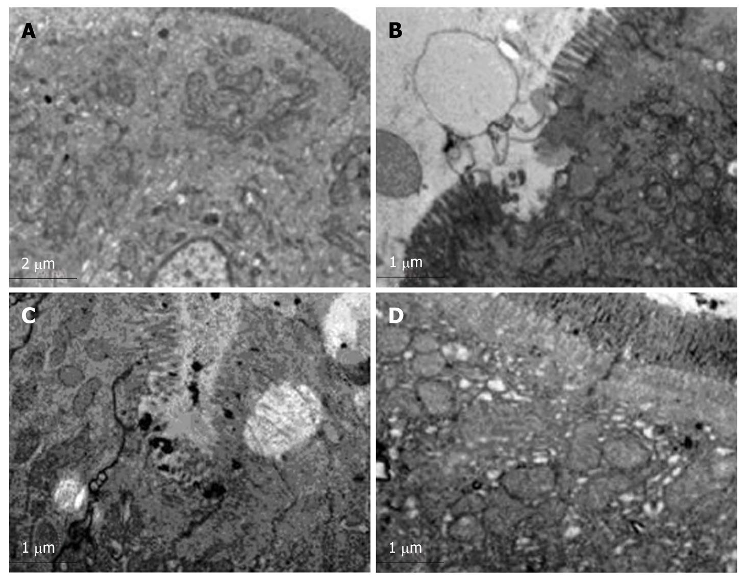
Figure 4 Transmission electron microscopy with nitric acid lanthanum tagging.
Orderly mucosal villi and integrated tight junctions as well as intact organelles with regular nuclei in group C (A), exfoliated and incomplete microvilli accompanying widened intercellular spaces as well as swollen endoplasmic reticulum and mitochondria and a small number of lanthanum granules in tissue spaces in group H (B), dilated Golgi complex with irregular nuclei and edge aggregation in chromatin as well as lanthanum granules in the tight junction gap and cells in group HH (C), mildly deformed microvilli and swollen mitochondria in lamina propria accompanying a small number of lanthanum granules confined to vessels and epithelial surface in group HG (D) (× 8900).
- Citation: Zhou QQ, Yang DZ, Luo YJ, Li SZ, Liu FY, Wang GS. Over-starvation aggravates intestinal injury and promotes bacterial and endotoxin translocation under high-altitude hypoxic environment. World J Gastroenterol 2011; 17(12): 1584-1593
- URL: https://www.wjgnet.com/1007-9327/full/v17/i12/1584.htm
- DOI: https://dx.doi.org/10.3748/wjg.v17.i12.1584


