Copyright
©2011 Baishideng Publishing Group Co.
World J Gastroenterol. Mar 21, 2011; 17(11): 1427-1433
Published online Mar 21, 2011. doi: 10.3748/wjg.v17.i11.1427
Published online Mar 21, 2011. doi: 10.3748/wjg.v17.i11.1427
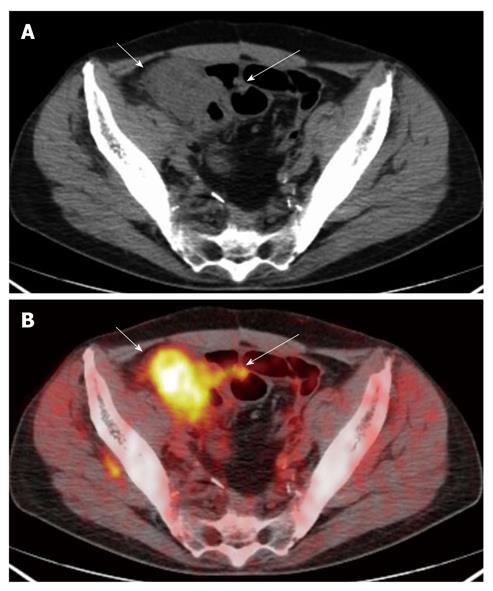
Figure 3 The computed tomography (A) and fused positron emission tomography/computed tomography (B) images show a neoplastic lesion of the caecum concentrating the radio-tracer (short arrows) (SUVmax 9.
6). Focal radio-tracer uptake (SUVmax 3.6) of a contiguous ileal loop was disclosed on positron emission tomography/computed tomography (B) (long arrow) consistent with a short concentric wall thickening (A) (long arrow): the finding was suggestive of small bowel infiltration. The lesion was correctly classified as T4.
- Citation: Mainenti PP, Iodice D, Segreto S, Storto G, Magliulo M, Palma GDD, Salvatore M, Pace L. Colorectal cancer and 18FDG-PET/CT: What about adding the T to the N parameter in loco-regional staging? World J Gastroenterol 2011; 17(11): 1427-1433
- URL: https://www.wjgnet.com/1007-9327/full/v17/i11/1427.htm
- DOI: https://dx.doi.org/10.3748/wjg.v17.i11.1427


