Copyright
©2011 Baishideng Publishing Group Co.
World J Gastroenterol. Mar 21, 2011; 17(11): 1383-1399
Published online Mar 21, 2011. doi: 10.3748/wjg.v17.i11.1383
Published online Mar 21, 2011. doi: 10.3748/wjg.v17.i11.1383
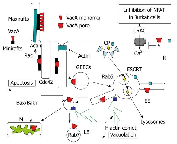
Figure 5 Endocytosis and intracellular trafficking of VacA.
Monomeric VacA may bind sphingomyelin on small rafts. Then, by formation of membrane extensions by actin polymerization via Rac activation, VacA is clustered in macrorafts where the p33 subunit oligomerizes and forms a channel that enters the membrane lipid bilayers. By a Cdc42-dependent process, VacA bound to sphingomelin associated to lipid rafts is transferred into cell membrane invaginations (tubules?), which are then pinched out from the membrane, with the help of F-actin filament, which leading to the formation of the glycosylphosphatidylinositol-anchored protein-enriched early endosomal compartment (GEEC) compartment that contains VacA. The toxin is then transferred to Rab5-positive early endosomes (EEs). In EEs, VacA is selectively addressed to EE tubular extensions that are formed by an F-actin process. These tubular extensions are pinched out from the EEs and form highly motile vacuoles that are propelled by F-actin comets. These motile vesicles then fuse with mitochondria (M) or late endosomes (LEs), where VacA induces apoptosis or vacuolation. The pro-apoptotic channels Bax and Bak may be brought to mitochondria by binding on VacA-containing motile vacuoles. Some VacA molecules may be recycled (R) back to the plasma membrane where the channel activity of the toxin, by altering the electric transmembrane potential, inhibits the voltage-dependent Ca2+ release-activated Ca2+ (CRAC) channel. This blocks entry of calcium that activates the calcineurin protease, which is required for processing of the nuclear factor (NFAT), and therefore inhibits the transcription of the interleukin 2 (IL-2) gene in Jurkat T-lymphocytes. Ligands entering the coated-pit pathway (CP) are directed, by the endosomal sorting complex required for transport (ESCRT) complex, towards internal vesicles of EEs and form the multivesicular body multivesicular body (MVB). MVBs are directed to lysosomes, by being propelled along microtubules, where the contents of EE internal vesicles are transferred and degraded.
- Citation: Ricci V, Romano M, Boquet P. Molecular cross-talk between Helicobacter pylori and human gastric mucosa. World J Gastroenterol 2011; 17(11): 1383-1399
- URL: https://www.wjgnet.com/1007-9327/full/v17/i11/1383.htm
- DOI: https://dx.doi.org/10.3748/wjg.v17.i11.1383


