Copyright
©2011 Baishideng Publishing Group Co.
World J Gastroenterol. Jan 7, 2011; 17(1): 69-78
Published online Jan 7, 2011. doi: 10.3748/wjg.v17.i1.69
Published online Jan 7, 2011. doi: 10.3748/wjg.v17.i1.69
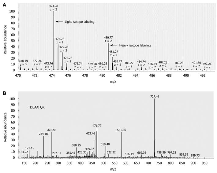
Figure 1 Identification of quantitatively dysregulated expression of S100A4.
A: Quantification of S100A4 through the isotopically labeled fragment ion signals of the peptide “TDEAAFQK”. The areas under the monoisotopic peaks represent the relative abundance of peptides, light [lymph node metastasis (LNM)]/heavy (non-LNM) = 3.04:1; B: Identification of the peptide “TDEAAFQK” from S100A4 by MS/MS.
- Citation: Huang LY, Xu Y, Cai GX, Guan ZQ, Sheng WQ, Lu HF, Xie LQ, Lu HJ, Cai SJ. S100A4 over-expression underlies lymph node metastasis and poor prognosis in colorectal cancer. World J Gastroenterol 2011; 17(1): 69-78
- URL: https://www.wjgnet.com/1007-9327/full/v17/i1/69.htm
- DOI: https://dx.doi.org/10.3748/wjg.v17.i1.69


