Copyright
©2011 Baishideng Publishing Group Co.
World J Gastroenterol. Jan 7, 2011; 17(1): 42-48
Published online Jan 7, 2011. doi: 10.3748/wjg.v17.i1.42
Published online Jan 7, 2011. doi: 10.3748/wjg.v17.i1.42
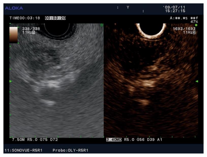
Figure 3 Contrast-enhanced (SonoVue) harmonic endoscopic ultrasound imaging showing a small (12 mm) hypovascular adenocarcinoma in the head of the pancreas.
The tumor tissue did not enhance in the early arterial phase, nor in the late venous phase, as compared to the surrounding pancreatic parenchyma.
- Citation: Reddy NK, Ioncică AM, Săftoiu A, Vilmann P, Bhutani MS. Contrast-enhanced endoscopic ultrasonography. World J Gastroenterol 2011; 17(1): 42-48
- URL: https://www.wjgnet.com/1007-9327/full/v17/i1/42.htm
- DOI: https://dx.doi.org/10.3748/wjg.v17.i1.42


