Copyright
©2011 Baishideng Publishing Group Co.
World J Gastroenterol. Jan 7, 2011; 17(1): 21-27
Published online Jan 7, 2011. doi: 10.3748/wjg.v17.i1.21
Published online Jan 7, 2011. doi: 10.3748/wjg.v17.i1.21
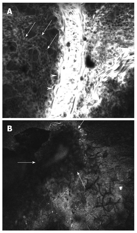
Figure 5 Confocal laser microscopy of the chick embryo chorioallantoic membrane.
A: Chick normal chorioallantoic membrane with visualization of large vessels (arrowheads), medium vessels (arrows) and circulating nucleated erythrocytes; B: Fragment of viable human colon cancer tissue (arrows) implanted on chick embryo chorioallantoic membrane.
- Citation: Gheonea DI, Cârţână T, Ciurea T, Popescu C, Bădărău A, Săftoiu A. Confocal laser endomicroscopy and immunoendoscopy for real-time assessment of vascularization in gastrointestinal malignancies. World J Gastroenterol 2011; 17(1): 21-27
- URL: https://www.wjgnet.com/1007-9327/full/v17/i1/21.htm
- DOI: https://dx.doi.org/10.3748/wjg.v17.i1.21


