Copyright
©2010 Baishideng.
World J Gastroenterol. Mar 7, 2010; 16(9): 1150-1154
Published online Mar 7, 2010. doi: 10.3748/wjg.v16.i9.1150
Published online Mar 7, 2010. doi: 10.3748/wjg.v16.i9.1150
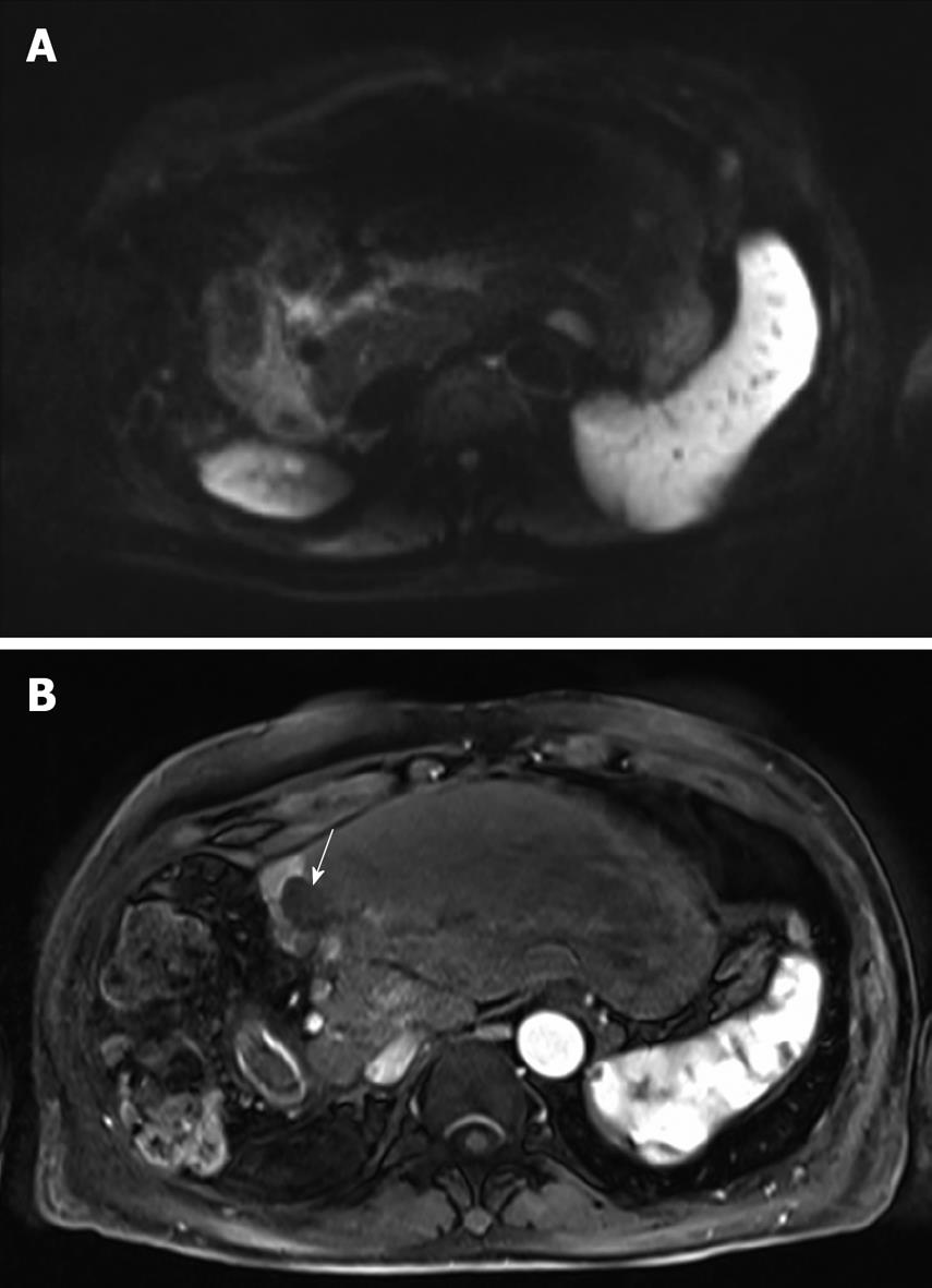
Figure 3 MRI of the liver after 8 cycles of HAI.
A: The lesion is isointense to the liver parenchyma on the diffusion-weighted image thereby suggesting the absence of viable tumor; B: The lesion is completely avascular (arrow) during the arterial phase after injection of gadolinium chelate. Note the enhancement of the remnant segment 4.
- Citation: Guiu B, Vincent J, Guiu S, Ladoire S, Ortega-Deballon P, Cercueil JP, Chauffert B, Ghiringhelli F. Hepatic arterial infusion of gemcitabine-oxaliplatin in a large metastasis from colon cancer. World J Gastroenterol 2010; 16(9): 1150-1154
- URL: https://www.wjgnet.com/1007-9327/full/v16/i9/1150.htm
- DOI: https://dx.doi.org/10.3748/wjg.v16.i9.1150


