Copyright
©2010 Baishideng.
World J Gastroenterol. Mar 7, 2010; 16(9): 1043-1049
Published online Mar 7, 2010. doi: 10.3748/wjg.v16.i9.1043
Published online Mar 7, 2010. doi: 10.3748/wjg.v16.i9.1043
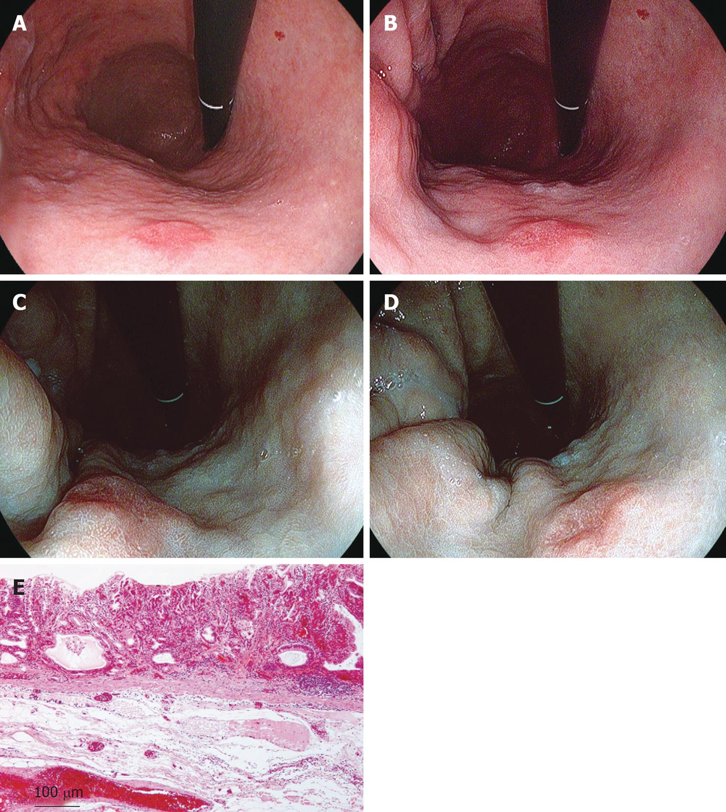
Figure 7 Case 2 (gastric cancer).
A: Conventional image: A red depressed lesions was visible on the anterior wall of the lower gastric body, but it was difficult to identify the demarcation of it; B: SE + CE image: The differences of the mucosal structure and color of the lesion could be observed; C, D: TE image: The demarcation of the lesion could be identified more clearly, observing changes in mucosal structure and color tone; E: Pathological diagnosis indicated a well to moderately differentiated adenocarcinoma, localized in the mucosal layer.
- Citation: Kodashima S, Fujishiro M. Novel image-enhanced endoscopy with i-scan technology. World J Gastroenterol 2010; 16(9): 1043-1049
- URL: https://www.wjgnet.com/1007-9327/full/v16/i9/1043.htm
- DOI: https://dx.doi.org/10.3748/wjg.v16.i9.1043


