Copyright
©2010 Baishideng.
World J Gastroenterol. Feb 28, 2010; 16(8): 1039-1042
Published online Feb 28, 2010. doi: 10.3748/wjg.v16.i8.1039
Published online Feb 28, 2010. doi: 10.3748/wjg.v16.i8.1039
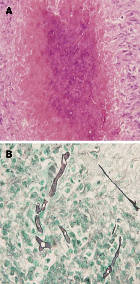
Figure 4 Pathological changes of liver tissue.
Inflammatory focus containing irregularly shaped, broad, nonseptate hyphae with right angle branching typical of mucormycosis. A: Hematoxylin and eosin stain, × 40; B: Grocott methenamine sliver stain, × 100.
- Citation: Li KW, Wen TF, Li GD. Hepatic mucormycosis mimicking hilar cholangiocarcinoma: A case report and literature review. World J Gastroenterol 2010; 16(8): 1039-1042
- URL: https://www.wjgnet.com/1007-9327/full/v16/i8/1039.htm
- DOI: https://dx.doi.org/10.3748/wjg.v16.i8.1039


