Copyright
©2010 Baishideng.
World J Gastroenterol. Feb 28, 2010; 16(8): 1034-1038
Published online Feb 28, 2010. doi: 10.3748/wjg.v16.i8.1034
Published online Feb 28, 2010. doi: 10.3748/wjg.v16.i8.1034
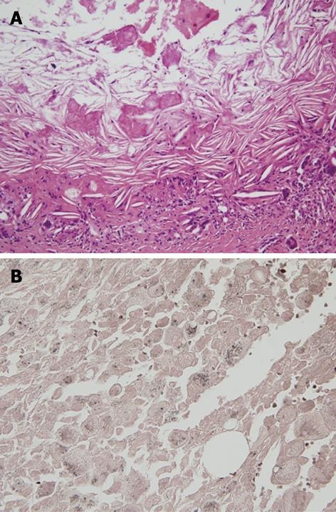
Figure 4 Microscopic finding of the pancreatic pseudocyst.
A: Microscopic finding of the pancreatic pseudocyst. HE staining showed multiple lipid droplets and cholesterol clefts; B: Sudan black B stain showed positive findings for lipid droplets, stained with a dark brown color.
- Citation: Cha SW, Kim SH, Lee HI, Lee YJ, Yang HW, Jung SH, Kim A, Lee MK, Han HY, Kang DW. Pancreatic pseudocyst filled with semisolid lipids mimicking solid mass on endoscopic ultrasound. World J Gastroenterol 2010; 16(8): 1034-1038
- URL: https://www.wjgnet.com/1007-9327/full/v16/i8/1034.htm
- DOI: https://dx.doi.org/10.3748/wjg.v16.i8.1034


