Copyright
©2010 Baishideng.
World J Gastroenterol. Feb 28, 2010; 16(8): 1034-1038
Published online Feb 28, 2010. doi: 10.3748/wjg.v16.i8.1034
Published online Feb 28, 2010. doi: 10.3748/wjg.v16.i8.1034
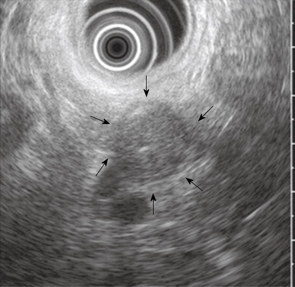
Figure 2 Image of the cystic lesion on EUS.
It showed a 17-mm × 19-mm, well-circumscribed, round, homogenous, hypoechoic solid lesion with central echogenicity (arrows).
- Citation: Cha SW, Kim SH, Lee HI, Lee YJ, Yang HW, Jung SH, Kim A, Lee MK, Han HY, Kang DW. Pancreatic pseudocyst filled with semisolid lipids mimicking solid mass on endoscopic ultrasound. World J Gastroenterol 2010; 16(8): 1034-1038
- URL: https://www.wjgnet.com/1007-9327/full/v16/i8/1034.htm
- DOI: https://dx.doi.org/10.3748/wjg.v16.i8.1034


