Copyright
©2010 Baishideng.
World J Gastroenterol. Feb 28, 2010; 16(8): 1003-1007
Published online Feb 28, 2010. doi: 10.3748/wjg.v16.i8.1003
Published online Feb 28, 2010. doi: 10.3748/wjg.v16.i8.1003
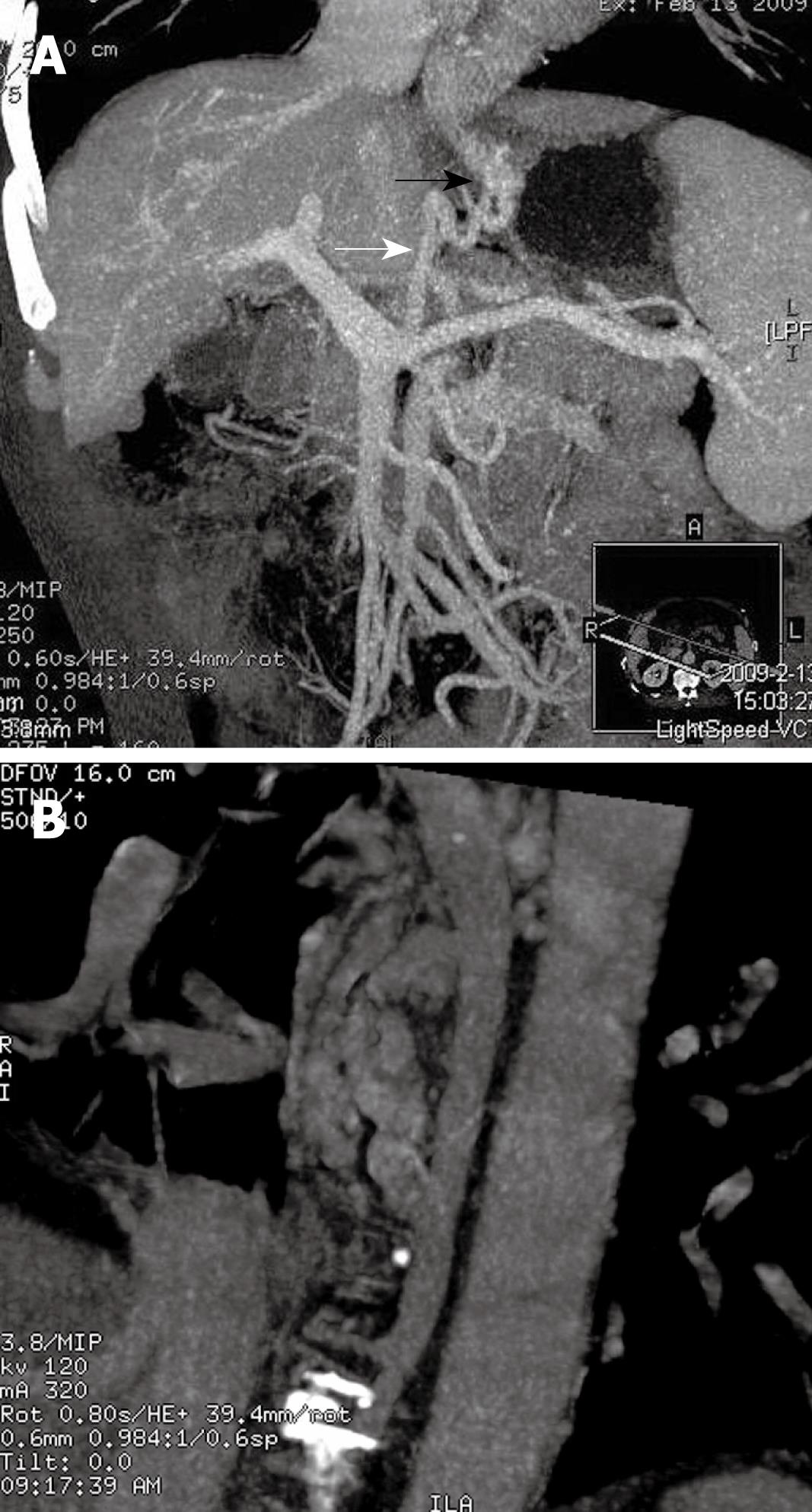
Figure 1 Gastroesophageal varices type 1.
A: Gastric varice (GV) (black arrow) originated from the left gastric vein (LGV) (white arrow); B: Venous drainage of the GV was via the azygos vein to the superior vena cava (arrow).
- Citation: Zhao LQ, He W, Ji M, Liu P, Li P. 64-row multidetector computed tomography portal venography of gastric variceal collateral circulation. World J Gastroenterol 2010; 16(8): 1003-1007
- URL: https://www.wjgnet.com/1007-9327/full/v16/i8/1003.htm
- DOI: https://dx.doi.org/10.3748/wjg.v16.i8.1003


