Copyright
©2010 Baishideng.
World J Gastroenterol. Feb 21, 2010; 16(7): 804-817
Published online Feb 21, 2010. doi: 10.3748/wjg.v16.i7.804
Published online Feb 21, 2010. doi: 10.3748/wjg.v16.i7.804
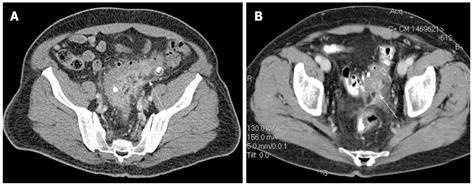
Figure 1 Diverticulitis.
A: Uncomplicated sigmoid diverticulitis with colonic thickening and straining at CT (arrow), also referred to as “mild” CT diverticulitis. Two diverticula contain contrast medium without evidence of extravasation outside the sigmoid; B: “Severe” CT diverticulitis with extravasation of contrast and small amount of extraluminal air (arrow). This patient was initially managed non-operatively and eventually required surgery for recurrent disease.
- Citation: Stocchi L. Current indications and role of surgery in the management of sigmoid diverticulitis. World J Gastroenterol 2010; 16(7): 804-817
- URL: https://www.wjgnet.com/1007-9327/full/v16/i7/804.htm
- DOI: https://dx.doi.org/10.3748/wjg.v16.i7.804


