Copyright
©2010 Baishideng.
World J Gastroenterol. Feb 14, 2010; 16(6): 723-727
Published online Feb 14, 2010. doi: 10.3748/wjg.v16.i6.723
Published online Feb 14, 2010. doi: 10.3748/wjg.v16.i6.723
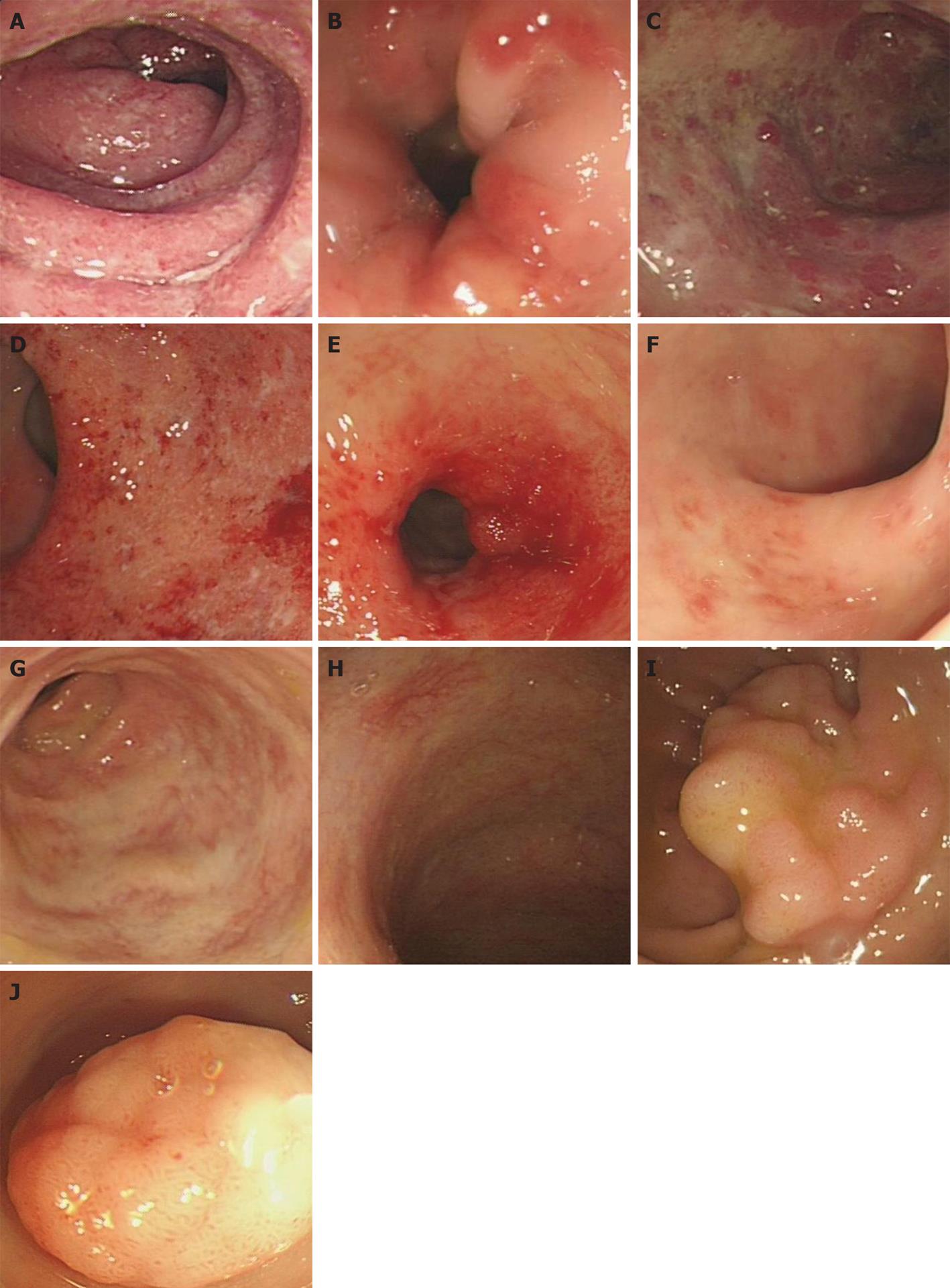
Figure 1 Endoscopic findings of schistosomal colonic disease.
A: Congestive, edematous mucosa in rectum with purulent secretion in mixed colitis; B: Congestive and edematous mucosa of sigmoid colon and intestinal stricture in mixed colitis; C: Mucosal erosion, superficial ulcer and granular change in descending colon with invisible submucosal blood vessels in mixed colitis; D: Congestive, edematous and erosive mucosa in rectum with invisible submucosal blood vessels in mixed colitis; E: Coarse, congestive, ulcerative mucosa and intestinal stricture in descending colon in mixed colitis; F: Patchy congestion and vague vascular net in mucosa of sigmoid colon in mixed colitis; G: Vascular net like map of sigmoid colon in chronic colitis; H: Cobwebbed vessels in rectum in chronic colitis; I: Giant flat, lobulated polypus in rectum in chronic colitis; J: Giant polypus in sigmoid colon in chronic colitis.
- Citation: Cao J, Liu WJ, Xu XY, Zou XP. Endoscopic findings and clinicopathologic characteristics of colonic schistosomiasis: A report of 46 cases. World J Gastroenterol 2010; 16(6): 723-727
- URL: https://www.wjgnet.com/1007-9327/full/v16/i6/723.htm
- DOI: https://dx.doi.org/10.3748/wjg.v16.i6.723


