Copyright
©2010 Baishideng.
World J Gastroenterol. Feb 14, 2010; 16(6): 713-722
Published online Feb 14, 2010. doi: 10.3748/wjg.v16.i6.713
Published online Feb 14, 2010. doi: 10.3748/wjg.v16.i6.713
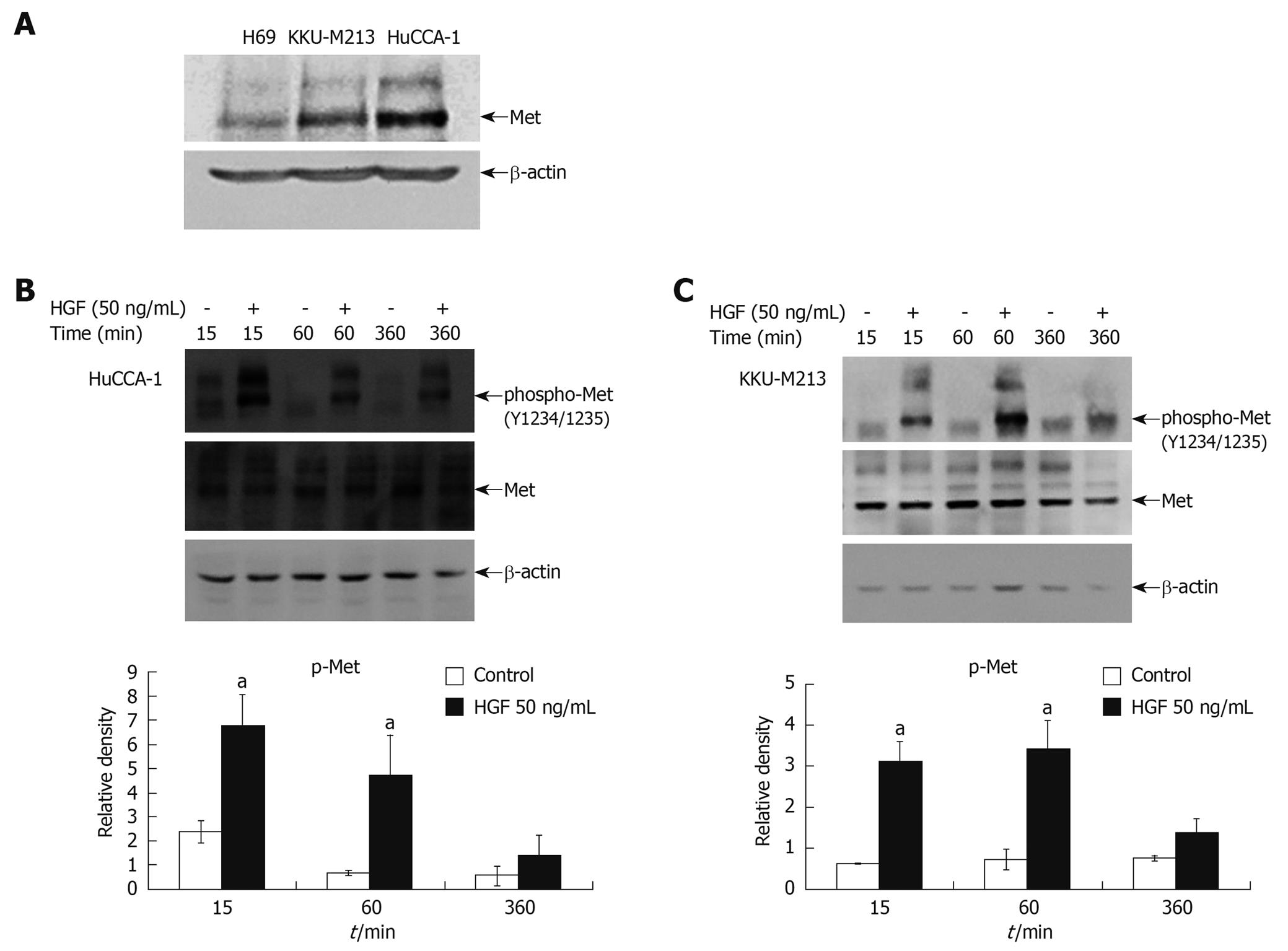
Figure 1 Steady state level of Met expression in cholangiocarcinoma cell lines and activation by hepatocyte growth factor (HGF).
Cell lysates from 80% confluent cells cultured in 10% fetal bovine serum (FBS) medium were examined for Met expression by Western blotting analysis (A). Lysates from HuCCA-1 (B) and KKU-M213 (C) cells treated with or without 50 ng/mL HGF for various times were analyzed by Western blotting for levels of Met and phospho-Met (pY1234/1235). The graphs show band densities of phospho-Met relative to those at zero time points. Data are presented as mean ± SE of results obtained from three independent experiments. aP < 0.05 vs untreated control.
- Citation: Menakongka A, Suthiphongchai T. Involvement of PI3K and ERK1/2 pathways in hepatocyte growth factor-induced cholangiocarcinoma cell invasion. World J Gastroenterol 2010; 16(6): 713-722
- URL: https://www.wjgnet.com/1007-9327/full/v16/i6/713.htm
- DOI: https://dx.doi.org/10.3748/wjg.v16.i6.713


