Copyright
©2010 Baishideng.
World J Gastroenterol. Feb 14, 2010; 16(6): 691-697
Published online Feb 14, 2010. doi: 10.3748/wjg.v16.i6.691
Published online Feb 14, 2010. doi: 10.3748/wjg.v16.i6.691
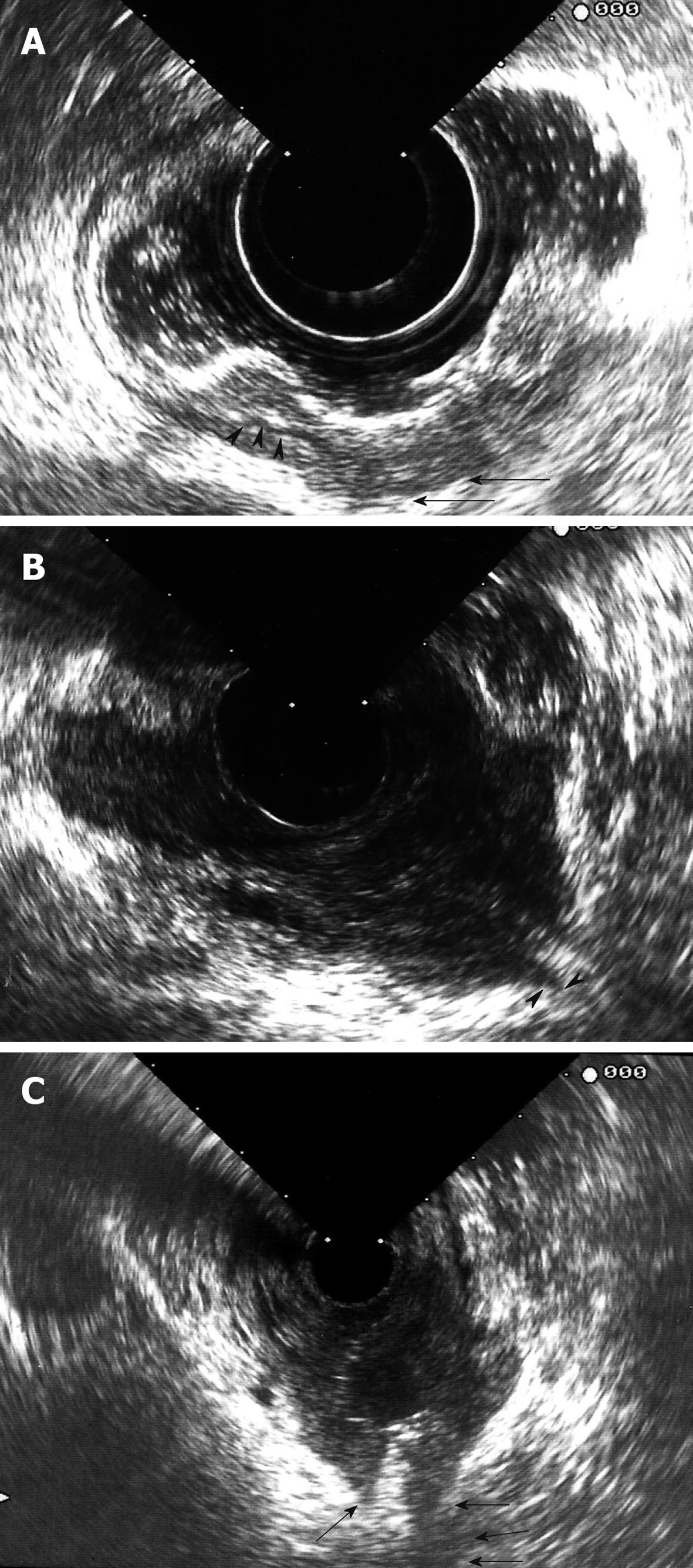
Figure 2 Endorectal ultrasound (ERUS) image.
A: A rectal carcinoma that appears to be T1 (penetration into submucosa) in one part (arrowheads show intact muscularis propria) and T2 (penetration into muscularis propria-arrows) in another part; B: A T3 rectal adenocarcinoma. Arrowheads show that the lesion penetrated into perirectal fat; C: A locally invasive cervical cancer, which invaded the rectum (arrows show tumor breach).
- Citation: Kav T, Bayraktar Y. How useful is rectal endosonography in the staging of rectal cancer? World J Gastroenterol 2010; 16(6): 691-697
- URL: https://www.wjgnet.com/1007-9327/full/v16/i6/691.htm
- DOI: https://dx.doi.org/10.3748/wjg.v16.i6.691


