Copyright
©2010 Baishideng.
World J Gastroenterol. Feb 14, 2010; 16(6): 691-697
Published online Feb 14, 2010. doi: 10.3748/wjg.v16.i6.691
Published online Feb 14, 2010. doi: 10.3748/wjg.v16.i6.691
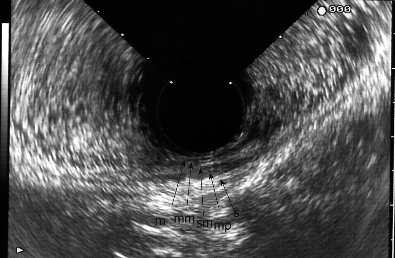
Figure 1 Normal endorectal sonogram image acquired by flexible echoendoscope.
The layers of the rectum are as follows: hyperechoic mucosa (m), hypoechoic muscularis mucosa (mm), hyperechoic submucosa (sm), hypoechoic muscularis propria (mp), and hyperechoic serosa (s).
- Citation: Kav T, Bayraktar Y. How useful is rectal endosonography in the staging of rectal cancer? World J Gastroenterol 2010; 16(6): 691-697
- URL: https://www.wjgnet.com/1007-9327/full/v16/i6/691.htm
- DOI: https://dx.doi.org/10.3748/wjg.v16.i6.691


