Copyright
©2010 Baishideng.
World J Gastroenterol. Feb 7, 2010; 16(5): 563-570
Published online Feb 7, 2010. doi: 10.3748/wjg.v16.i5.563
Published online Feb 7, 2010. doi: 10.3748/wjg.v16.i5.563
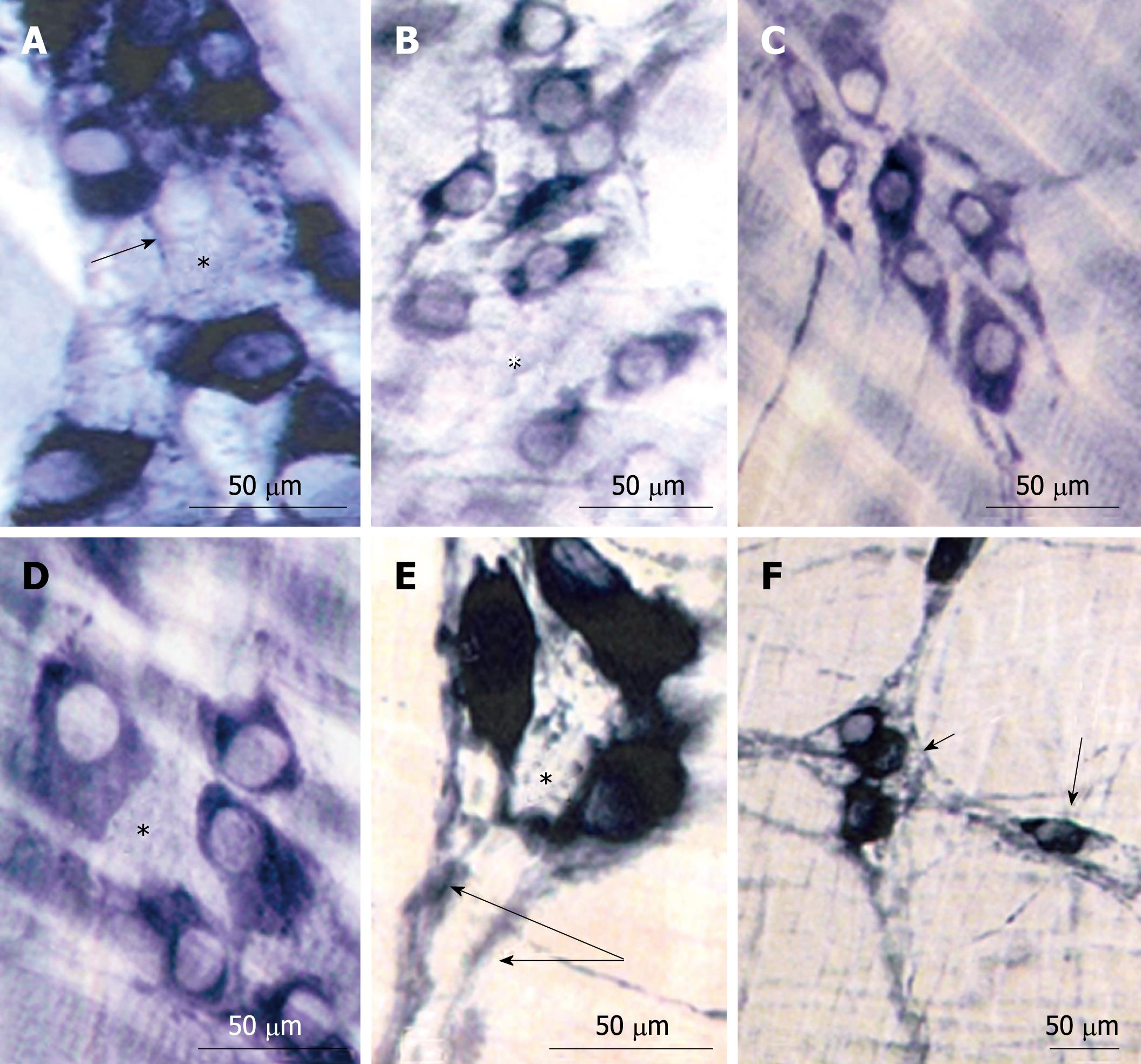
Figure 2 NADPH¨Cdiaphorase reaction.
A and D: N42 group. Note that most of the myenteric neurons were intensely reactive and spaces inside the ganglia (*) are surrounded by thin neuronal branches (arrow); B and E: D42 group. Neurons with diverse intensities of reaction, spaces inside the ganglia (*) and thick neuronal meshes (double-arrow) were evident; C and F: R42 group. Neuronal meshes surrounded the ganglion (small arrow). Some neurons were detected inside the meshes (large arrow).
- Citation: Greggio FM, Fontes RB, Maifrino LB, Castelucci P, Souza RR, Liberti EA. Effects of perinatal protein deprivation and recovery on esophageal myenteric plexus. World J Gastroenterol 2010; 16(5): 563-570
- URL: https://www.wjgnet.com/1007-9327/full/v16/i5/563.htm
- DOI: https://dx.doi.org/10.3748/wjg.v16.i5.563


