Copyright
©2010 Baishideng.
World J Gastroenterol. Feb 7, 2010; 16(5): 563-570
Published online Feb 7, 2010. doi: 10.3748/wjg.v16.i5.563
Published online Feb 7, 2010. doi: 10.3748/wjg.v16.i5.563
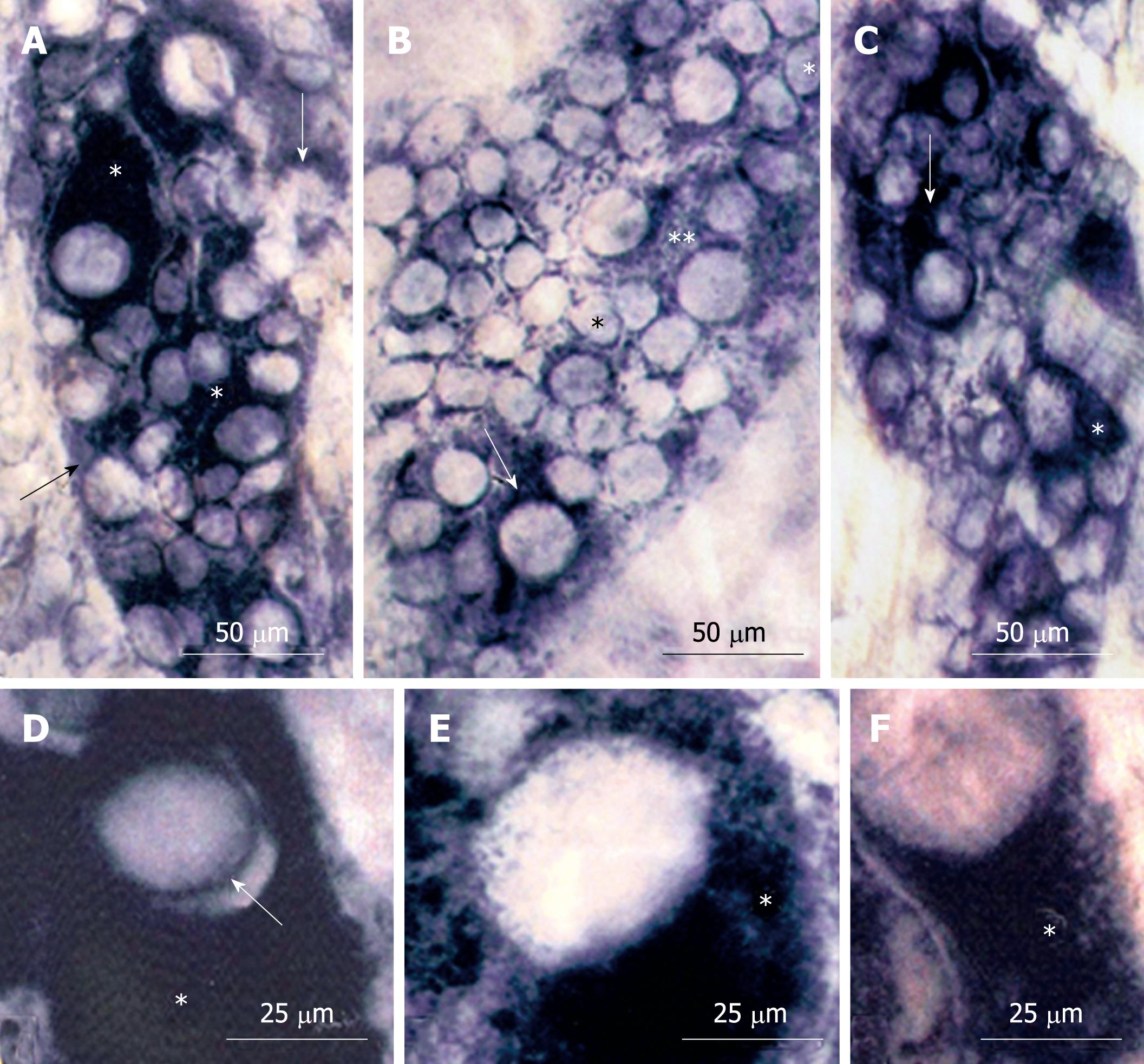
Figure 1 NADH-diaphorase reaction.
A: N42 group. Myenteric neurons of large and medium sizes with intensely reactive cytoplasm (*) and neurons of diverse size weakly reactive (arrows); B: D42 group. Nuclei of small neurons (*). The cytoplasm of large neurons had low (**) or diffuse (arrow) reactivity; C: R42 group. Note large neurons with intense (arrow) and diffuse (*) cytoplasmic reactivity; D-F: Large neurons, respectively from N42 (D), D42 (E) and R42 (F) groups. The well-delineated nucleus (arrow) from N42 was not clearly detected in malnourished and protein-recovered animals. Compare the patterns of reactivity of the cytoplasm (*).
- Citation: Greggio FM, Fontes RB, Maifrino LB, Castelucci P, Souza RR, Liberti EA. Effects of perinatal protein deprivation and recovery on esophageal myenteric plexus. World J Gastroenterol 2010; 16(5): 563-570
- URL: https://www.wjgnet.com/1007-9327/full/v16/i5/563.htm
- DOI: https://dx.doi.org/10.3748/wjg.v16.i5.563


