Copyright
©2010 Baishideng Publishing Group Co.
World J Gastroenterol. Dec 14, 2010; 16(46): 5822-5829
Published online Dec 14, 2010. doi: 10.3748/wjg.v16.i46.5822
Published online Dec 14, 2010. doi: 10.3748/wjg.v16.i46.5822
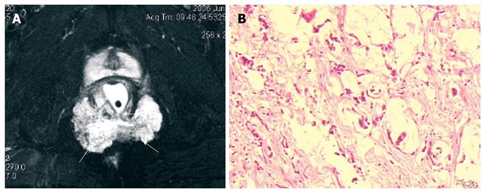
Figure 5 A 59-year-old man with mucinous adenocarcinoma caused by anal fistula.
A: Axial fat-suppressed T2-weighted magnetic resonance image shows a horse-shoe mass with typical mesh-like enhancing areas (arrows). The mass has a internal fistula connected to the anorectum (arrows); B: Microscopy shows a single or small cluster of atypical cells floating in mucin pool and bundles of collagen with hyaline degeneration in stroma.
- Citation: Yang BL, Gu YF, Shao WJ, Chen HJ, Sun GD, Jin HY, Zhu X. Retrorectal tumors in adults: Magnetic resonance imaging findings. World J Gastroenterol 2010; 16(46): 5822-5829
- URL: https://www.wjgnet.com/1007-9327/full/v16/i46/5822.htm
- DOI: https://dx.doi.org/10.3748/wjg.v16.i46.5822


