Copyright
©2010 Baishideng Publishing Group Co.
World J Gastroenterol. Dec 7, 2010; 16(45): 5766-5772
Published online Dec 7, 2010. doi: 10.3748/wjg.v16.i45.5766
Published online Dec 7, 2010. doi: 10.3748/wjg.v16.i45.5766
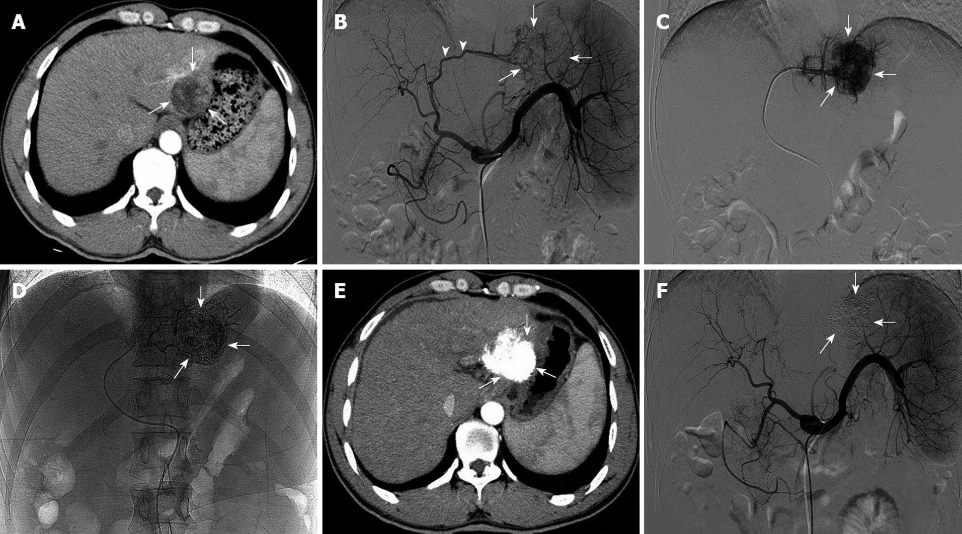
Figure 1 A newly diagnosed patient with single-nodule treated by transarterial embolization ablation.
A: An enhanced computed tomography (CT) scan of hepatocellular carcinoma tumor before treatment. The tumor measured 3.5 cm × 3.3 cm (arrows); B: Hepatic artery angiography showed the thick blood vessels of the tumor (arrowheads) and the abnormal vascular group (arrows) before treatment; C: Superselective angiography clearly showed tumor staining (arrows); D: After transarterial embolization ablation with an lipiodol-ethanol mixture, lipiodol accumulated in the tumor (arrows); E: An enhanced CT scan 2 mo after treatment showed dense deposition of lipiodol in the tumor without enhancement (arrows); F: Hepatic artery angiography showed the absence of tumor blood vessels and tumor staining at 2 mo after treatment (arrows).
- Citation: Gu YK, Luo RG, Huang JH, Si Tu QJ, Li XX, Gao F. Transarterial embolization ablation of hepatocellular carcinoma with a lipiodol-ethanol mixture. World J Gastroenterol 2010; 16(45): 5766-5772
- URL: https://www.wjgnet.com/1007-9327/full/v16/i45/5766.htm
- DOI: https://dx.doi.org/10.3748/wjg.v16.i45.5766


