Copyright
©2010 Baishideng Publishing Group Co.
World J Gastroenterol. Nov 28, 2010; 16(44): 5582-5587
Published online Nov 28, 2010. doi: 10.3748/wjg.v16.i44.5582
Published online Nov 28, 2010. doi: 10.3748/wjg.v16.i44.5582
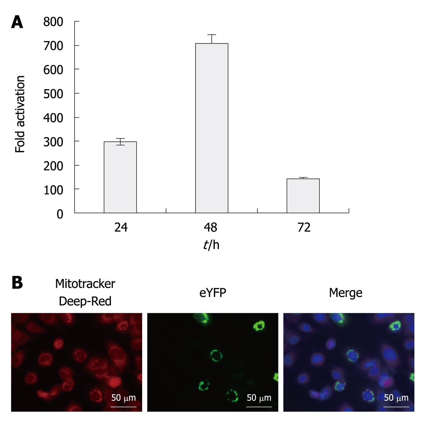
Figure 1 Activation of the interferon-β promoter by enhanced yellow fluorescent protein-mitochondrial antiviral signaling protein and subcellular localization of enhanced yellow fluorescent protein-mitochondrial antiviral signaling protein.
A: Activation of the interferon (IFN)-β promoter by enhanced yellow fluorescent protein (eYFP)-mitochondrial antiviral signaling protein (MAVS). Expression vector of eYFP-MAVS was co-transfected with IFN-β-secreted placental alkaline phosphatase (SEAP) in Huh7.5 cells. pRL-TK was co-transfected to normalize transfection efficiency. SEAP activity in cell culture was measured at 24, 48 and 72 h post-transfection. Results are expressed as activation levels of the promoter compared to those in cells transfected with an empty expression vector. The error bars represent the SDs from the mean values obtained from three independent experiments performed in duplicate; B: Fluorescence microscopy of Huh7.5 cells transfected with eYFP-MAVS at 48 h post-transfection. Mitochondria were stained with Mitotracker deep red (red) and nuclei were labeled with 4’,6-Diamidino-2-phenylindole (blue). Yellow labeling in the merged image indicates co-localization of eYFP-MAVS with mitochondria.
- Citation: Fu QX, Wang LC, Jia SZ, Gao B, Zhou Y, Du J, Wang YL, Wang XH, Peng JC, Zhan LS. Screening compounds against HCV based on MAVS/IFN-β pathway in a replicon model. World J Gastroenterol 2010; 16(44): 5582-5587
- URL: https://www.wjgnet.com/1007-9327/full/v16/i44/5582.htm
- DOI: https://dx.doi.org/10.3748/wjg.v16.i44.5582


