Copyright
©2010 Baishideng Publishing Group Co.
World J Gastroenterol. Nov 21, 2010; 16(43): 5474-5480
Published online Nov 21, 2010. doi: 10.3748/wjg.v16.i43.5474
Published online Nov 21, 2010. doi: 10.3748/wjg.v16.i43.5474
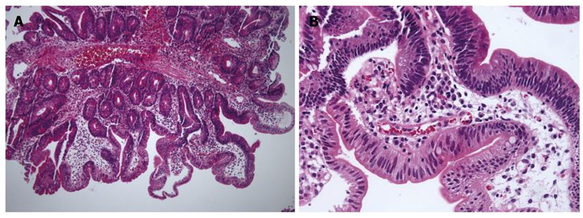
Figure 5 Microscopic finding of serrated adenomas.
A: Vascular stalk and saw-tooth appearance were observed (HE, × 40); B: At high magnification, hyperplastic foveolar cells were found. In part, epithelia with pleomorphic, stratified nuclei and irregular chromatin deposits were observed (HE, × 200).
- Citation: Jung SH, Chung WC, Kim EJ, Kim SH, Paik CN, Lee BI, Cho YS, Lee KM. Evaluation of non-ampullary duodenal polyps: Comparison of non-neoplastic and neoplastic lesions. World J Gastroenterol 2010; 16(43): 5474-5480
- URL: https://www.wjgnet.com/1007-9327/full/v16/i43/5474.htm
- DOI: https://dx.doi.org/10.3748/wjg.v16.i43.5474


