Copyright
©2010 Baishideng Publishing Group Co.
World J Gastroenterol. Nov 7, 2010; 16(41): 5263-5266
Published online Nov 7, 2010. doi: 10.3748/wjg.v16.i41.5263
Published online Nov 7, 2010. doi: 10.3748/wjg.v16.i41.5263
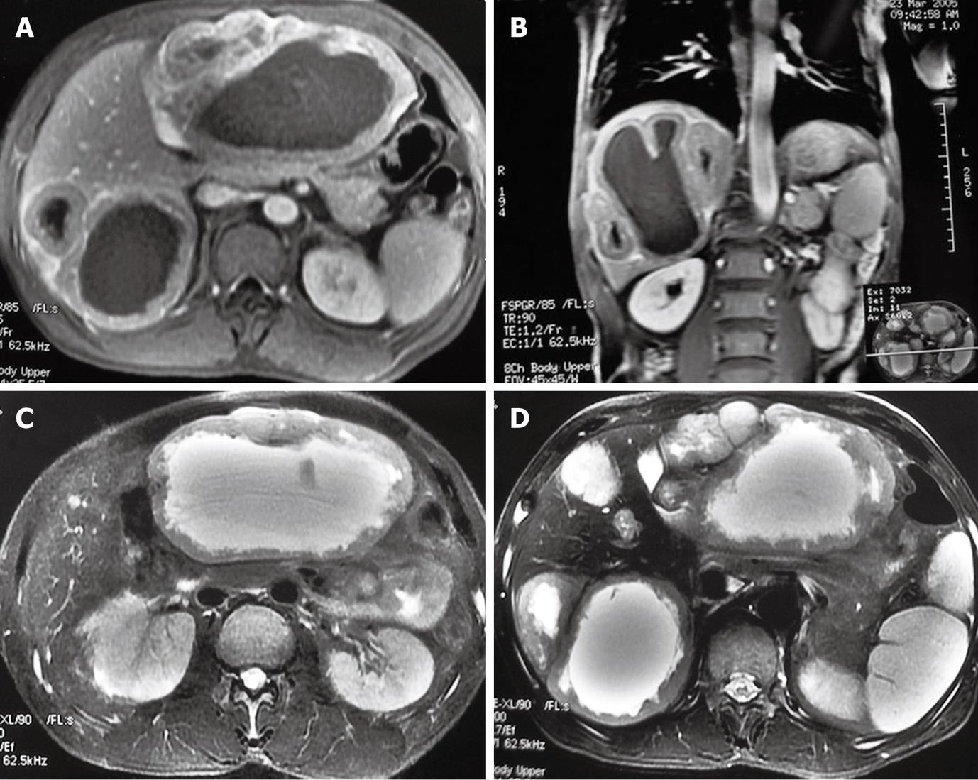
Figure 1 Axial fat-suppressed T2-weighted turbo spin echo magnetic resonance imaging showing two large lesions (A) and several small cyst-like lesions (B) in liver, and coronal and transverse T1-weighted imaging showing thickened wall of lesions with a ragged appearance (C) and enhancement (D) in liver.
- Citation: Chen J, Du YJ, Song JT, E LN, Liu BR. Primary malignant liver mesenchymal tumor: A case report. World J Gastroenterol 2010; 16(41): 5263-5266
- URL: https://www.wjgnet.com/1007-9327/full/v16/i41/5263.htm
- DOI: https://dx.doi.org/10.3748/wjg.v16.i41.5263


