Copyright
©2010 Baishideng Publishing Group Co.
World J Gastroenterol. Oct 28, 2010; 16(40): 5092-5103
Published online Oct 28, 2010. doi: 10.3748/wjg.v16.i40.5092
Published online Oct 28, 2010. doi: 10.3748/wjg.v16.i40.5092
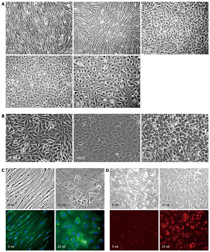
Figure 1 Hepatic differentiation of human bone marrow-derived mesenchymal stem cells.
A: Morphological characterization of differentiated mesenchymal stem cells (MSCs) under hepatic induction. 0 wk: Undifferentiated MSCs; 1 wk: 1 wk post-induction; 4 wk: 4 wk post-induction; 7 wk: 7 wk post-induction; and 10 wk: 10 wk post-induction (original magnification, × 50); B: Morphology of human hepatoma cell lines: SK-Hep-1, HA22T/VGH, and HepG2 (original magnification, × 100); C: Production of albumin (green color) in differentiated MSCs after 10 wk induction (10 wk) and counterstained with Hoechst (blue color). The undifferentiated MSCs (0 wk) were used as negative control cells (original magnification, × 200); D: Uptake of low-density lipoprotein (red color) in differentiated MSCs after 10 wk induction (10 wk). The undifferentiated MSCs (0 wk) were used as negative control cells (original magnification, × 100).
- Citation: Chen ML, Lee KD, Huang HC, Tsai YL, Wu YC, Kuo TM, Hu CP, Chang C. HNF-4α determines hepatic differentiation of human mesenchymal stem cells from bone marrow. World J Gastroenterol 2010; 16(40): 5092-5103
- URL: https://www.wjgnet.com/1007-9327/full/v16/i40/5092.htm
- DOI: https://dx.doi.org/10.3748/wjg.v16.i40.5092


