Copyright
©2010 Baishideng Publishing Group Co.
World J Gastroenterol. Oct 28, 2010; 16(40): 5016-5019
Published online Oct 28, 2010. doi: 10.3748/wjg.v16.i40.5016
Published online Oct 28, 2010. doi: 10.3748/wjg.v16.i40.5016
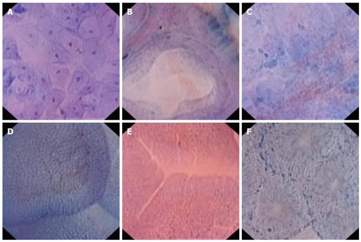
Figure 1 The various endocytoscopic images seen in the gastrointestinal tract.
A: Normal esophageal mucosa on endocytoscopy (EC): Squamous cells can be seen with nuclei that have a regular shape and size. There is good nuclei:cytoplasm ratio; B: Normal Barrett’s mucosa on EC: Regular glandular structure can be observed with homogenous intestinal metaplasia cells containing uniform appearing nucleus bordering the glands; C: EC in Barrett’s esophagus with intramucosal cancer: There is total loss of the glandular pattern and markedly increased cellular density. The nuclei appear pleomorphic and enlarged; D: EC in normal mucosa in the duodenum: The villi can be clearly visualized with presence of regular appearing villous capillaries. Numerous nuclei can also be seen within each villi; E: In a patient with Marsh 3 biopsy-proven celiac disease, the mucosa appears atropic with complete absence of any villous structure. There is a “cracked mud” appearance and capillaries are distinctly absent in contrast to the normal pattern; F: EC performed on normal colonic mucosa reveals uniform appearing glands which are regularly arranged. Each gland is lined by epithelium and they are arranged in a radial fashion with the colonic crypt seen centrally.
- Citation: Singh R, Mei SLCY, Tam W, Raju D, Ruszkiewicz A. Real-time histology with the endocytoscope. World J Gastroenterol 2010; 16(40): 5016-5019
- URL: https://www.wjgnet.com/1007-9327/full/v16/i40/5016.htm
- DOI: https://dx.doi.org/10.3748/wjg.v16.i40.5016


