Copyright
©2010 Baishideng.
World J Gastroenterol. Jan 28, 2010; 16(4): 508-512
Published online Jan 28, 2010. doi: 10.3748/wjg.v16.i4.508
Published online Jan 28, 2010. doi: 10.3748/wjg.v16.i4.508
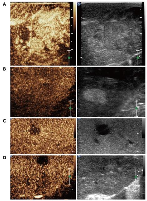
Figure 2 CE-IOUS showing an HCC nodule with hyperenhancement in arterial phase (A, arrow) while a regenerative hyperechoic nodule shows isoenhancement on CE-IOUS (B, arrow); Hypoechoic intrahepatic metastatic nodule showing wash out of contrast agent on late phase (C, arrow); Isoechoic nodule missed on IOUS showing a clear margin on CE-IOUS (D, arrow).
- Citation: Wu H, Lu Q, Luo Y, He XL, Zeng Y. Application of contrast-enhanced intraoperative ultrasonography in the decision-making about hepatocellular carcinoma operation. World J Gastroenterol 2010; 16(4): 508-512
- URL: https://www.wjgnet.com/1007-9327/full/v16/i4/508.htm
- DOI: https://dx.doi.org/10.3748/wjg.v16.i4.508


