Copyright
copy;2010 Baishideng Publishing Group Co.
World J Gastroenterol. Oct 21, 2010; 16(39): 4998-5004
Published online Oct 21, 2010. doi: 10.3748/wjg.v16.i39.4998
Published online Oct 21, 2010. doi: 10.3748/wjg.v16.i39.4998
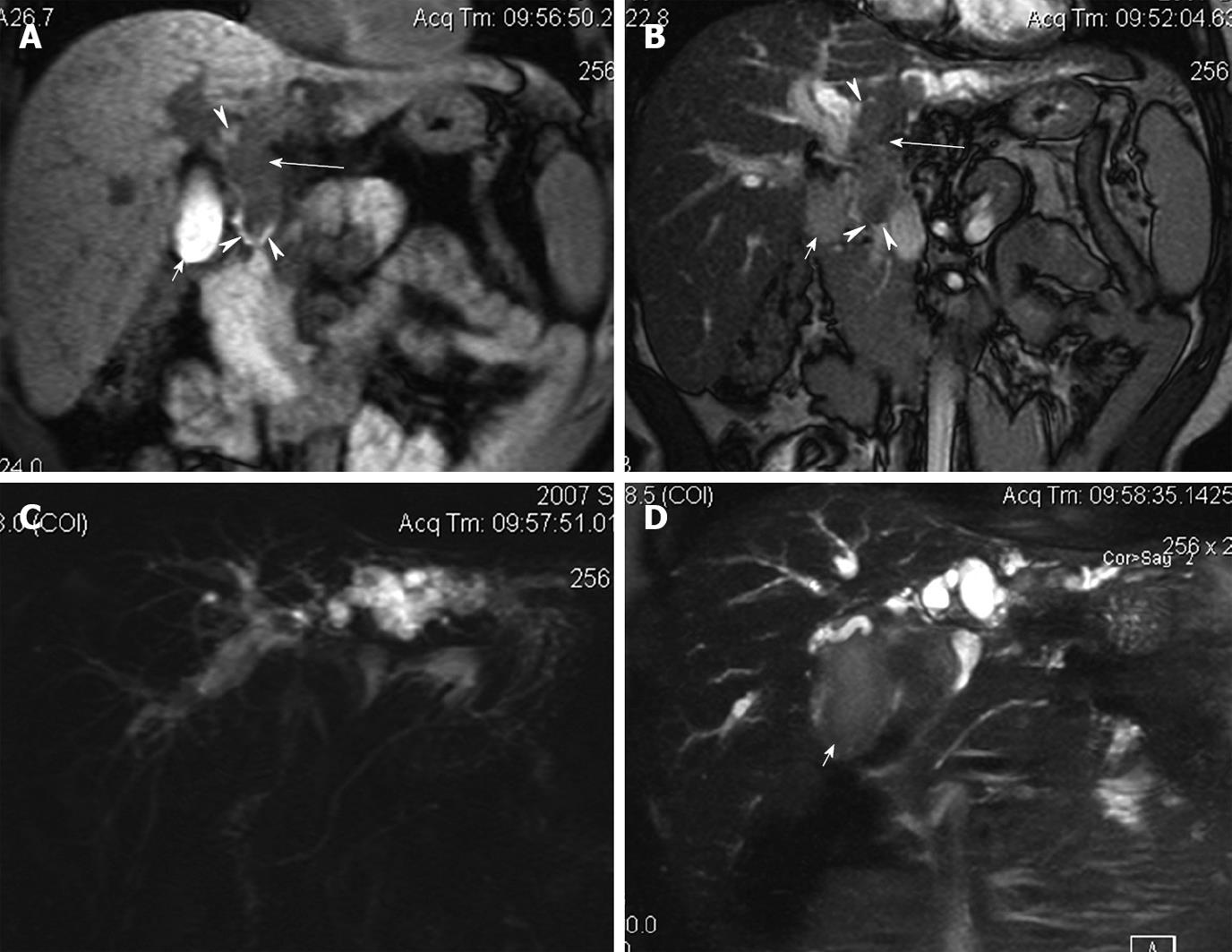
Figure 2 Magnetic resonance and magnetic resonance cholangiopancreatography images of Case 2.
A: Coronal T1 weighted image showing hypo-intense tumor thrombus (large arrows) in left hepatic, common hepatic and common bile ducts; B: Coronal T2 weighted image showing the tumor thrombus as iso- to slight hyper-intense; C: Thick slice magnetic resonance cholangiopancreatography image showing bilateral dilated intrahepatic ducts with “vanished” common hepatic and common bile ducts; D: Thin slice magnetic resonance cholangiopancreatography image showing filling defect in the hilar. Neither portal vein thrombus nor hepatic parenchymal mass is identified. The signal intensity of liver is heterogeneous due to cirrhosis. Blood clots or hemorrhage can be observed in gallbladder (small arrows), common hepatic and common bile ducts (arrowheads), which are hyper-dense on T1 weighted image, and slight hyper-intense on T2 weighted image.
- Citation: Long XY, Li YX, Wu W, Li L, Cao J. Diagnosis of bile duct hepatocellular carcinoma thrombus without obvious intrahepatic mass. World J Gastroenterol 2010; 16(39): 4998-5004
- URL: https://www.wjgnet.com/1007-9327/full/v16/i39/4998.htm
- DOI: https://dx.doi.org/10.3748/wjg.v16.i39.4998


