Copyright
copy;2010 Baishideng Publishing Group Co.
World J Gastroenterol. Oct 21, 2010; 16(39): 4938-4943
Published online Oct 21, 2010. doi: 10.3748/wjg.v16.i39.4938
Published online Oct 21, 2010. doi: 10.3748/wjg.v16.i39.4938
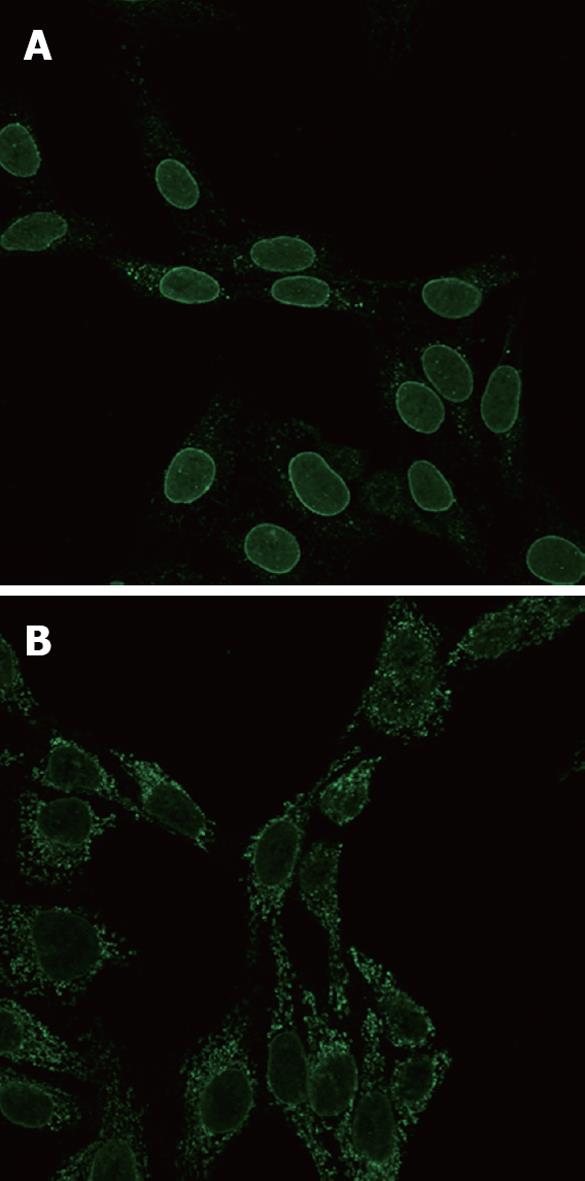
Figure 1 Typical peri-nuclear staining showing anti-nuclear envelope antibody positive sera in indirect immunofluorescence.
A: Cells fixed with 1% formaldehyde; B: Cells fixed with 4% formaldehyde.
- Citation: Sfakianaki O, Koulentaki M, Tzardi M, Tsangaridou E, Theodoropoulos PA, Castanas E, Kouroumalis EA. Peri-nuclear antibodies correlate with survival in Greek primary biliary cirrhosis patients. World J Gastroenterol 2010; 16(39): 4938-4943
- URL: https://www.wjgnet.com/1007-9327/full/v16/i39/4938.htm
- DOI: https://dx.doi.org/10.3748/wjg.v16.i39.4938


