Copyright
©2010 Baishideng Publishing Group Co.
World J Gastroenterol. Oct 7, 2010; 16(37): 4725-4732
Published online Oct 7, 2010. doi: 10.3748/wjg.v16.i37.4725
Published online Oct 7, 2010. doi: 10.3748/wjg.v16.i37.4725
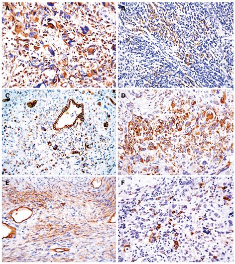
Figure 3 Immunohistochemistry showing tumor cells strongly reactive to vimentin (A), diffuse membranous immunostaining for CD56 in mesenchymal cells (B), diffuse multifocal cytoplasmic immunostaining with a distinct paranuclear dot-like staining using cytokeratin 19 (C), focal cytoplasmic positivity for desmin in some tumor cells (D), tumor cells focally positive for α-smooth muscle actin (E) and S100 (F) (EnVision+, × 400).
- Citation: Li XW, Gong SJ, Song WH, Zhu JJ, Pan CH, Wu MC, Xu AM. Undifferentiated liver embryonal sarcoma in adults: A report of four cases and literature review. World J Gastroenterol 2010; 16(37): 4725-4732
- URL: https://www.wjgnet.com/1007-9327/full/v16/i37/4725.htm
- DOI: https://dx.doi.org/10.3748/wjg.v16.i37.4725


