Copyright
©2010 Baishideng Publishing Group Co.
World J Gastroenterol. Oct 7, 2010; 16(37): 4725-4732
Published online Oct 7, 2010. doi: 10.3748/wjg.v16.i37.4725
Published online Oct 7, 2010. doi: 10.3748/wjg.v16.i37.4725
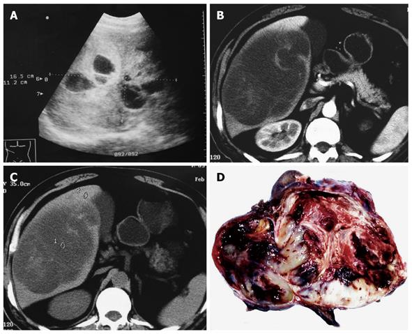
Figure 1 Abdominal ultrasonography showing a 16.
5 cm × 11.2 cm multilocular cystic liver mass (A), computed tomography imaging demonstrating a large, hypodense tumor occupying the right lobe of liver with multicystic (B) and solid portions (C), and polychromatic cut surface which is soft with fluid and mucoid zones, firm with fleshy areas and necrotico-hemorrhagic changes (D).
- Citation: Li XW, Gong SJ, Song WH, Zhu JJ, Pan CH, Wu MC, Xu AM. Undifferentiated liver embryonal sarcoma in adults: A report of four cases and literature review. World J Gastroenterol 2010; 16(37): 4725-4732
- URL: https://www.wjgnet.com/1007-9327/full/v16/i37/4725.htm
- DOI: https://dx.doi.org/10.3748/wjg.v16.i37.4725


