Copyright
©2010 Baishideng Publishing Group Co.
World J Gastroenterol. Oct 7, 2010; 16(37): 4634-4639
Published online Oct 7, 2010. doi: 10.3748/wjg.v16.i37.4634
Published online Oct 7, 2010. doi: 10.3748/wjg.v16.i37.4634
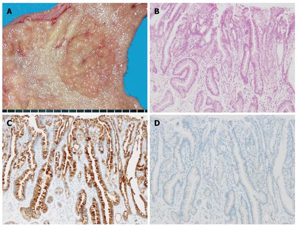
Figure 3 Gastric-type differentiated adenocarcinoma.
A: Macroscopic appearance, showing a fine-granule aggregated lesion; B: Well-differentiated tubular adenocarcinoma (HE staining); C: Diffuse positive staining of MUC5AC was apparent in the carcinomatous gland; D: No staining of MUC2 was evident.
- Citation: Namikawa T, Hanazaki K. Mucin phenotype of gastric cancer and clinicopathology of gastric-type differentiated adenocarcinoma. World J Gastroenterol 2010; 16(37): 4634-4639
- URL: https://www.wjgnet.com/1007-9327/full/v16/i37/4634.htm
- DOI: https://dx.doi.org/10.3748/wjg.v16.i37.4634


